LDH-Glo™ Cytotoxicity Assay


Simple, More Sensitive LDH Assay Method to Determine Cytotoxicity
- Detects LDH release from small numbers of cells, including 3D cell models
- Monitors cytotoxicity from the same sample over time
- Provides more data per well through multiplexing with other cell-based assays
- Integrates seamlessly with the MyGlo® Reagent Reader

Catalog Number:
Choose a product
Size
Catalog Number: J2380
Catalog Number: J2381
少数細胞での細胞毒性モニタリング
LDH-Glo™ Cytotoxicity Assay は細胞膜損傷により培地に放出される乳酸脱水素酵素 (LDH) をプレートベースで発光測定します。発光による測定は発色法や蛍光法に比べて高感度検出を可能にするため、少数の細胞 (初代培養細胞や3D細胞培養を含む)から漏出する LDH でも正確に検出することができます。
このアッセイでは処理後の各ウェルより極少量の培地 (2–5µl) を使用するだけなので、長時間にわたり同じウェル中のサンプルからより多くのデーターを取得できることができ、残った培地や細胞を利用して他の細胞ベースアッセイを行うこともできます。
【簡易マニュアル】日本語
※日本語マニュアルは英語版を翻訳したものですが、常に更新されるものではありません。最新のマニュアルについては以下の ”Complete Protocol” をご覧ください。
How Does the LDH-Glo™ Cytotoxicity Assay Work?
アッセイの原理
ダメージを受けた細胞から漏出した LDH は、NAD+ から NADH への還元を伴い乳酸の酸化を触媒します。レダクターゼは NADHと基質を用いてルシフェリンを生成し、これが Ultra-Glo™ rLuciferase により生物発光シグナルに変換されます。
How Do I Monitor Cytotoxicity in 3D Cell Cultures?
The LDH-Glo™ Cytotoxicity Assay is well suited for measuring LDH released from small numbers of membrane-damaged cells. Here is an example of measuring drug-induced toxicity in HCT116 spheroids. The time-dependent toxicity measurements were performed by repeatedly sampling media from the same wells.
How do I run the LDH-Glo™ assay with 3D spheroids or organoids, and what controls should I include?
Remove just 2–5µl of medium per timepoint—up to about 10% of a well's starting volume doesn't measurably affect remaining cells or spheroids—dilute into LDH Storage Buffer, and freeze or assay directly. Every plate should include a No-Cell Control (medium background), a Vehicle-Only Control, and, if calculating percent cytotoxicity, a Maximum LDH Release Control using Triton™ X-100.
Because 3D models can have heterogeneous baseline LDH release, normalize to a same-plate, same-treatment-day control rather than comparing raw luminescence values across separate experiments.

How Do I Detect Target Cell Cytotoxicity in ADCC Assays?
ADCC アッセイでのターゲット細胞の細胞毒性検出
LDH-Glo™ Cytotoxicity assay は 補体依存性細胞傷害 (CDC)、抗体依存性細胞媒介性細胞傷害 (ADCC) や抗体-薬物複合体の細胞毒性などバイオロジクス処理の作用機序にともなう様々な細胞死の評価に使用することができます。生物製剤 リツキシマブ を用いたターゲット細胞の ADCC細胞死の検出にLDH-Glo™ Assayを用いた例を示します。

Pair with the MyGlo® Reagent Reader
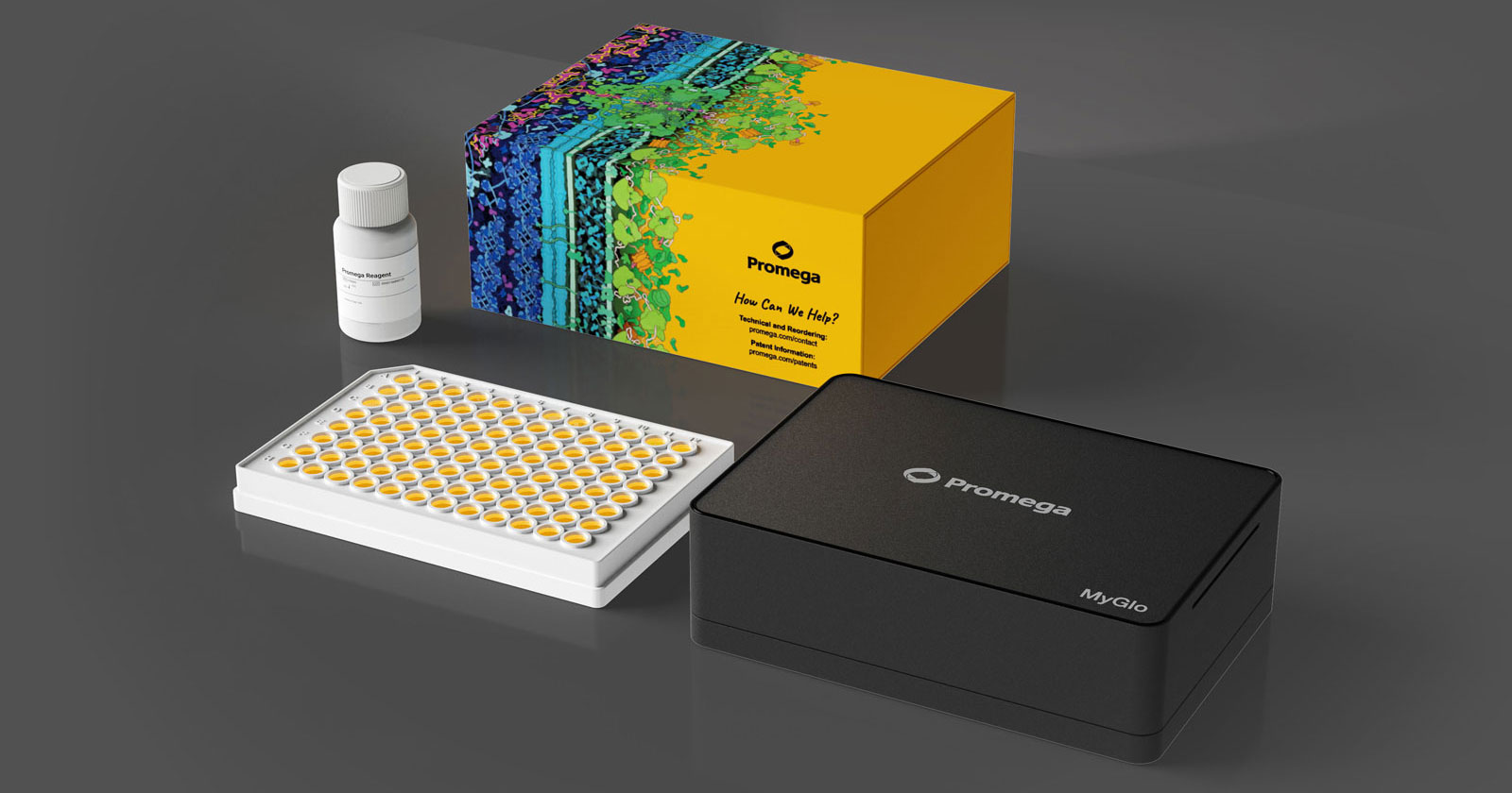
Reading your assay results doesn't require a shared plate reader. The MyGlo® Reagent Reader is a compact 96-well luminescence plate reader, enabling you to run luminescent assays at your bench without competing for instrument time. Integrated software generates dose response curves, single concentration and linearity analysis in minutes.
Protocols
Complete Protocol
Frequently Asked Questions
How can the LDH-Glo™ assay use only 2–5µl of medium for repeated sampling?
Because bioluminescent detection is highly sensitive, only a very small sample is needed to generate a measurable signal. Removing 2–5µl—up to roughly 10% of a well's starting medium volume across several timepoints—doesn't measurably affect the remaining cells, so the same well can be sampled repeatedly to build a time-course without disturbing the culture.
What controls are recommended for LDH-Glo™ assay in 3D cultures?
Every assay plate should include a No-Cell Control (triplicate wells without cells, to measure medium background) and a Vehicle-Only Cells Control (untreated cells with the same solvent used for test compounds). If you need percent cytotoxicity, add a Maximum LDH Release Control by treating vehicle-only cells with Triton™ X-100 to lyse them completely before sampling.
Can LDH-Glo™ assay samples be frozen and measured later?
Yes. Samples frozen in LDH Storage Buffer show no significant loss of LDH activity after a freeze-thaw cycle, but samples frozen directly in PBS with BSA can lose up to 85% of LDH activity—so always store in LDH Storage Buffer before freezing, not PBS.
Can the LDH-Glo™ assay be multiplexed with a cell viability assay?
Yes. Because only a small volume of medium is removed for LDH-Glo™ detection, the remaining sample can be used for RealTime-Glo™ MT, CellTiter-Glo® or CellTiter-Fluor™ viability measurements, or for orthogonal endpoints like Caspase-Glo® 3/7, Glucose-Glo™ or Glutamate-Glo™ assays—helping distinguish cytostatic from cytotoxic effects in the same well.

How do you choose the right sample dilution factor for the LDH-Glo™ assay?
Dilution depends on cell number and whether serum is present, since serum itself contains LDH. Promega's technical manual recommends roughly 5-fold dilution for under 1,000 cells/100µl without serum, up to 100-fold for 10,000–50,000 cells/100µl without serum, and up to 300-fold with 10% serum at higher cell numbers—always confirm results fall within the assay's linear range.
Why is bioluminescent LDH detection more sensitive at low signal levels than colorimetric detection?
Bioluminescence produces light against an essentially dark background, so even small amounts of signal are easy to distinguish from noise. Colorimetric detection instead measures a small color change against a comparatively bright background (transmitted light), which requires more analyte to produce a reliably measurable difference—making it less suited to low-cell-number or low-LDH samples.
Specifications
Catalog Number:
選択製品の構成品内容
| Item | Part # | Size |
|---|---|---|
Reductase Substrate |
G885A | 1 × 55μl |
Lactate Dehydrogenase |
J195A | 1 × 1 each |
LDH Detection Enzyme Mix |
J238A | 1 × 10ml |

U.S. Pat. Nos. 9,273,343 and 9,951,372, European Pat. No. 2751089, Japanese Pat. No. 6067019 and other patents pending.
選択製品の構成品内容
| Item | Part # | Size |
|---|---|---|
Reductase Substrate |
G885B | 1 × 275μl |
Lactate Dehydrogenase |
J195A | 1 × 1 each |
LDH Detection Enzyme Mix |
J238B | 1 × 50ml |
U.S. Pat. Nos. 9,273,343 and 9,951,372, European Pat. No. 2751089, Japanese Pat. No. 6067019 and other patents pending.
Related Products
類似製品
CellTox™ Green Cytotoxicity Assay
細胞死に伴う細胞膜破壊により細胞毒性を蛍光で測定
G8741, G8742, G8743, G8731
CytoTox-Glo™ Cytotoxicity Assay
細胞集団中の死細胞数を発光で測定する細胞毒性アッセイ
G9290, G9291, G9292
CytoTox-ONE™ Homogeneous Membrane Integrity Assay
死細胞を測定するホモジニアスなLDH ベースの蛍光アッセイ
G7890, G7891, G7892
CytoTox-Fluor™ Cytotoxicity Assay
培養中の死細胞数を測定する蛍光アッセイ
G9260, G9261, G9262
関連製品
CellTiter-Glo® 3D Cell Viability Assay
ATP量測定をベースに三次元培養中の生細胞数をホモジニアスな発光法により定量
G9681, G9682, G9683
GloMax® Discover System
発光・蛍光・吸光度測定に対応した高性能マイクロプレートリーダー
GM3000
RealTime-Glo™ MT Cell Viability Assay
72 時間まで培養細胞の生存性をモニタリングできる発光アッセイ
G9711, G9712, G9713
Caspase-Glo® 3/7 Assay System
カスパーゼ-3/7活性の検出に対応した、使いやすいプレートベースの発光アッセイ
G8090, G8091, G8093, G8092
Resources
技術記事
- "Will This Drug Cause Hepatotoxicity?"
- App Note: Predicting gastrointestinal toxicity
- Impact of aerosols on liver xenobiotic metabolism: A comparison of two methods of exposure
- Folate and Vitamin B12 Deficiency Exacerbate Inflammation during Mycobacterium avium paratuberculosis (MAP) Infection.
- SARS‐CoV‐2‐reactive T‐cell receptors isolated from convalescent COVID‐19 patients confer potent T‐cell effector function