3D細胞培養における生物学的変化をモニタリングするためのアッセイとツール
- 3D細胞培養では、細胞の健全性、代謝、生細胞タンパク質ダイナミクス、遺伝子発現解析、ゲノム変化など、モニタリングすべき重要なパラメーターが複数あります。
- アッセイの選択基準:1)生物学的な問い、2)溶解系 vs. 非溶解系、3)マルチプレックス解析の必要性、4)下流のアプリケーション
- 非溶解アッセイは下流解析のために3D培養を保持できます。一方、溶解アッセイは細胞を溶解しながらスフェロイド内部まで試薬を浸透させます。
- 同一ウェルでのマルチプレックス解析により、限られた3Dサンプルから最大限のデータ取得が可能です。細胞生存性+細胞毒性+発現測定を組み合わせることが可能です。
- プロメガのアッセイの多くはプロトコルを変更せずに3Dで使用できます。スフェロイドやオルガノイドへの浸透には、一部プロトコルの最適化が必要な場合があります。
新しいアプローチ方法論(New Approach Methodologies; NAMs)では現在、再現性、経時的な追跡、複雑なモデル間での標準化について、FDAによる明確なバリデーション要件が求められています。スフェロイド、オルガノイド、臓器チップを中心としたワークフローを構築している場合、これらの要件は使用するすべてのアッセイに及びます。プロメガは20年以上にわたり、こうした課題に対応し、NAMs研究の要求に応えるための生物発光アッセイの開発に取り組んできました。
3D細胞培養を使用する理由
研究者たちは今日、細胞生物学を研究するためのモデルシステムを構築するにあたり、スフェロイド、オルガノイド、臓器チップなどの3D培養法を活用するようになっています。3D細胞培養は、マトリックスとのより自然な相互作用を促進し、細胞が独自のマトリックスを分泌することも可能にすることから、2D環境よりも組織や腫瘍の環境を忠実に再現します。また、より自然な細胞間接触を提供し、酸素や栄養素へのアクセスに勾配が生じます。 3D細胞培養は、単層培養と比較して、細胞応答において顕著な差異を明らかにすることがあります。3D培養における細胞の生物学的変化のモニタリングは、細胞の健全性、代謝、発現の変化を観察することで行えます。これらの表現型の変化は、最終的には細胞の遺伝子型、あるいはその変化が疾患状態を引き起こした遺伝子型の変異と関連づけられます。プロメガは、細胞の健全性や代謝変化から遺伝子発現・解析まで、ワークフローの各ステップに対応するアッセイとツールを提供しています。
3D細胞培養の概要、使用目的と方法、3Dモデルに適したアッセイの選び方について説明しています。
3D細胞培養における細胞の健全性はどのようにモニタリングできますか?
セルヘルスアッセイは、細胞生存性、細胞毒性、アポトーシスを測定します。大半は単層培養向けに設計されていますが、3Dモデルではプロトコルの最適化が必要です。スフェロイドやオルガノイドの中心部まで試薬を浸透させ、細胞マーカーを測定用に放出させることが課題となります。プロメガは、溶解アッセイ(完全溶解・高シグナル)と非溶解アッセイ(下流解析のために細胞を保存)の両方を、3D対応で提供しています。
3D細胞培養における生物学的変化のモニタリングが初めての方へ
3D培養モデルを使用する際には、適切なアッセイシステムの選択が重要です。3D培養における生物学的変化のモニタリングに使用できるツールについてご紹介します。
CellTiter-Glo® 3D Cell Viability Assay
溶解アッセイ
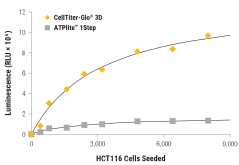
3D培養に特化して設計された試薬によるATP測定で、細胞生存性をモニタリングします。従来のCellTiter-Glo®アッセイと同じATP測定ルシフェラーゼケミストリーを採用しながら、より高い溶解力により深部への浸透を実現し、定量に必要なATPをより多く放出できます。CellTox™ Green Cytotoxicity Assayとの同一ウェルでのマルチプレックス解析、またはLDH-Glo™ Cytotoxicity Assayによる培地サンプリングで、死細胞の同時測定が可能です。
単一オルガノイドを用いた創薬スクリーニング
オルガノイドは、ヒト組織内の細胞不均一性をより忠実に再現することから、創薬スクリーニングにおけるin vivo応答の予測精度を高める上で不可欠なツールです。このようなモデルの創薬スクリーニングへの需要は高まっているものの、再現性やスケーラビリティの欠如といったワークフロー上の課題から、オルガノイドを用いたin vitroアッセイのHTSへの適用はまだ限られています。Cell MicrosystemsとプロメガはここでHTS用プレートベースアッセイに最適化・検証された、数百個のオルガノイドを再現性よく自動作製するソリューションを紹介します。
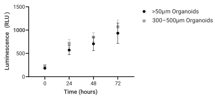
未選別オルガノイドと選別オルガノイドの細胞生存性比較
CellTiter-Glo® 3D Cell Viability Assay
溶解アッセイ
3D培養に特化して設計された試薬によるATP測定で、細胞生存性をモニタリングします。従来のCellTiter-Glo®アッセイと同じATP測定ルシフェラーゼケミストリーを採用しながら、より高い溶解力により深部への浸透を実現し、定量に必要なATPをより多く放出できます。CellTox™ Green Cytotoxicity Assayとの同一ウェルでのマルチプレックス解析、またはLDH-Glo™ Cytotoxicity Assayによる培地サンプリングで、死細胞の同時測定が可能です。
CellTiter-Glo® 3DとATPlite 1Step試薬の溶解能の比較
RealTime-Glo™ MT Cell Viability Assay
非溶解アッセイ
生物発光アッセイにより、最長72時間にわたって継続的に細胞生存性をモニタリングします。細胞内の還元当量を検出する高感度なアッセイです。同一ウェルから遺伝子発現またはゲノム解析用の核酸を抽出することが可能です。CellTox™ Greenとのマルチプレックス解析で死細胞の測定にも対応します。
HCT116スフェロイドの直径増加に対するRealTime-Glo™ MT Cell Viability Assayの応答
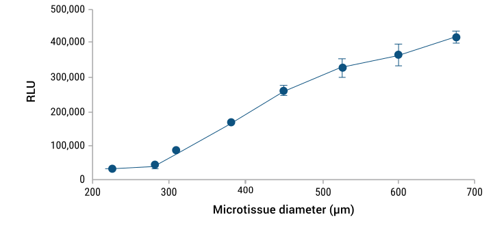
詳細はアプリケーションノート(下記リンク)をご参照ください。
CellTox™ Green Cytotoxicity Assay
非溶解アッセイ
蛍光アッセイにより、最長72時間にわたって継続的に細胞毒性をモニタリングします。細胞非透過性のDNA結合色素が、膜が損傷した細胞に侵入して細胞DNAを染色します。RealTime-Glo™ MTとのマルチプレックス解析で生細胞・死細胞の継続モニタリングが可能です。また、CellTiter-Glo® 3Dのエンドポイント解析に先行して使用することもできます。
生細胞・死細胞モニタリングのための同一ウェルマルチプレックス解析
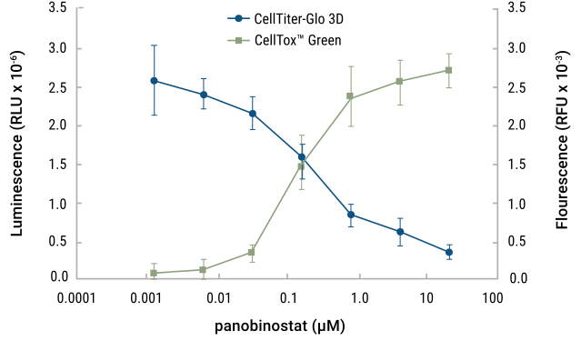
詳細はサイエンティフィックポスター(下記リンク)をご参照ください。
LDH-Glo™ Cytotoxicity Assay
非溶解アッセイ
高感度な生物発光アッセイにより、乳酸脱水素酵素(LDH)の放出を指標として細胞毒性を評価します。1タイムポイントあたり培地2〜5μlのみで測定可能なため、同一ウェルから複数回のサンプリングが行えます。CellTiter-Glo® 3DやCaspase-Glo® 3/7 Assayなど、その後のセルベースアッセイに先行して細胞毒性を測定する際にご使用ください。
LDH-Glo™アッセイとCellTiter-Glo®アッセイのマルチプレックス解析
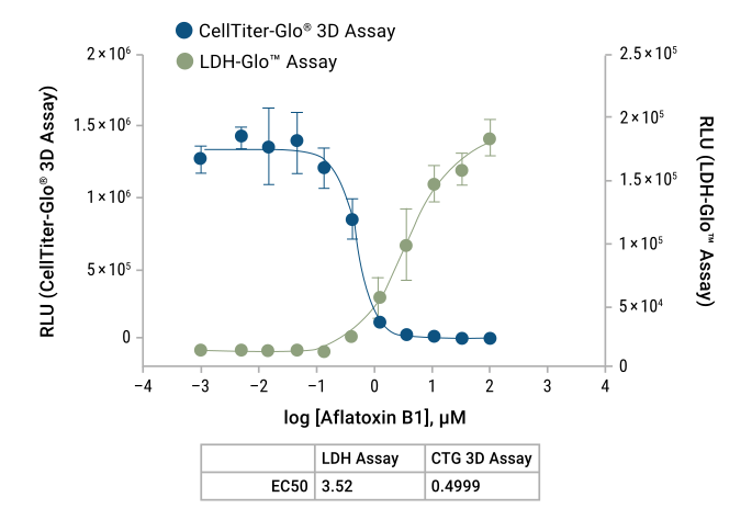
ヒト肝臓マイクロティッシュスフェロイドをアフラトキシンB1で48時間処理した。培地サンプルを採取し、LDH-Glo™アッセイで測定した。残存細胞はCellTiter-Glo® 3Dアッセイで細胞生存性を測定した。詳細はテクニカルマニュアル TM548を参照。
Caspase-Glo® 3/7 3D Assay
溶解アッセイ
カスパーゼ-3および-7活性を測定する高感度な生物発光アッセイで、アポトーシスをモニタリングします。細胞毒性アッセイおよび細胞生存性アッセイとのマルチプレックス解析で、アポトーシス応答をより包括的に評価できます。
CellTox™ Green Cytotoxicity Assay、CellTiter-Fluor™ Cell Viability AssayおよびCaspase-Glo® 3/7 3D Assayのマルチプレックス解析
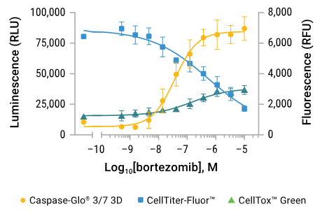
A549スフェロイドにボルテゾミブを24時間暴露した後、同一ウェルでマルチプレックス測定を実施した。蛍光および発光はGloMax® Discover装置で測定した。
RealTime-Glo™ Annexin V Apoptosis and Necrosis Assay
非溶解アッセイ
マルチプレックス化された生物発光アネキシンVアッセイと蛍光ネクローシスアッセイにより、アポトーシスと二次ネクローシスを最大48時間まで連続的にモニタリングできます。同じウェルから核酸を単離し、遺伝子発現解析やゲノム解析に使用できます。
HepG2スフェロイドにおけるアネキシンV露出および二次ネクローシスの測定
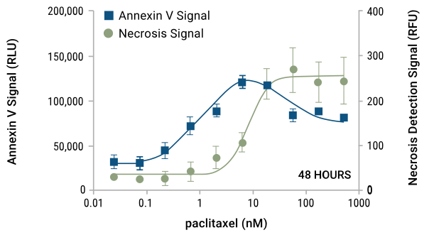
RealTime-Glo™ Annexin V Apoptosis and Necrosis Assayを使用し、パクリタキセルを48時間暴露したHepG2スフェロイドのアネキシンV露出および二次ネクローシスを測定した。発光および蛍光はGloMax® Discoverで測定した。
P450‐Glo™ Assays
非溶解アッセイ(オプション)
生物発光アッセイにより、シトクロムP450酵素(CYP1A2、 CYP2B6、CYP2C9 )の活性化をモニタリングします。非溶解プロトコルオプションでは、単層培養と3D培養で同じプロトコルを使用します。プロ基質が細胞内に拡散し、CYP活性によってルシフェリンに変換された後、培地中に拡散したルシフェリンをアッセイします。細胞はその後も他のアッセイや核酸抽出に使用できます。
ヒト肝臓マイクロティッシュのCYP3A4活性と細胞生存性のモニタリング
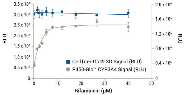
リファンピシン応答におけるヒト肝臓マイクロティッシュのシトクロムP450 3A4活性と細胞生存性をモニタリングした。CYP3A4活性は非溶解P450-Glo™ 3A4 Assay(Luciferin-IPA使用)で測定し、続いて同一ウェルでCellTiter-Glo® 3D Assayにより細胞生存性を測定した。測定にはGloMax® Discoverを用いた。
Autophagy LC3 HiBiT Reporter Assay System
非溶解アッセイ
プレートベースの生物発光アッセイで、オートファジー性LC3-IIフラックスをモニタリングします。LC3 HiBiTレポーターを安定発現させた細胞を3D培養し、Lytic HiBiT Detection Systemを用いてモニタリングします。このレポーターは、蛍光顕微鏡を用いたLC3-IIタンパク質のモニタリングにも使用できます。
LC3 HiBiTレポーター発現HEK293スフェロイドのオートファジー刺激剤および阻害剤に対する応答
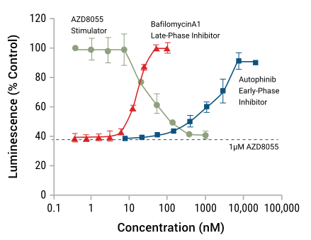
詳細はサイエンティフィックポスター(下記リンク)をご参照ください。
RealTime-Glo™ Extracellular ATP Assay
非溶解アッセイ
スタウロスポリン処理したHCT-116スフェロイドからのATP放出
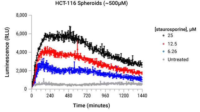
RealTime-Glo™ Extracellular ATP Assayを用いてATP放出をモニタリングし、24時間にわたって10分ごとに測定した。
Lumit® Cell Proliferation Assay (Human Ki-67)
溶解アッセイ
細胞増殖の広く知られたマーカーであるhKi-67を、洗浄ステップ不要で早い時点から追跡できる、添加・測定形式のプレートベースアッセイです。キットにはCellTox™ Green色素が含まれており、同一ウェルで細胞毒性と増殖のマルチプレックス検出が可能です。
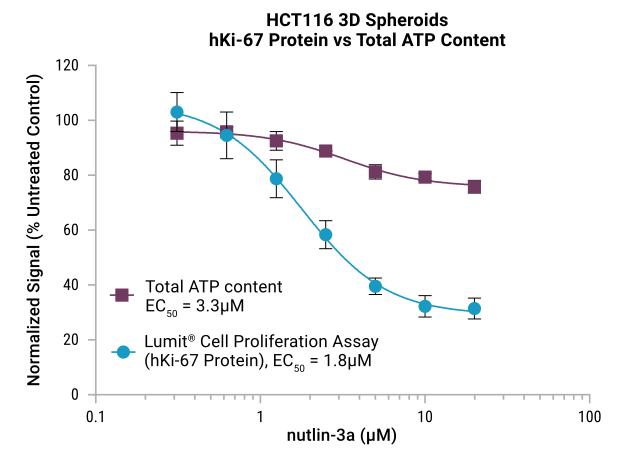
図:HCT116細胞(1,000細胞/ウェル)をSBio PrimeSurface 96Uプレート(ULA;丸底)に播種し、3日間培養して3Dスフェロイドを形成した。得られたHCT116スフェロイドをnutlin-3aの濃度を変えて24時間処理した。その後、増殖をLumit® Cell Proliferation Assay(Human Ki-67)で、代謝活性を別プレートで評価した。
3D細胞培養に対応する代謝アッセイ
エネルギー代謝は細胞の健康状態と機能にとって重要であり、代謝産物は細胞エネルギー、細胞構成要素の合成、シグナル伝達経路と密接に関連しています。プロメガは、3D細胞培養アプリケーション向けに最適化されたプロトコルを備えた、代謝活性を測定する複数のアッセイを提供しています。非溶解アッセイオプションでは、同一スフェロイドから複数タイムポイントにわたって測定が可能です。
注目製品
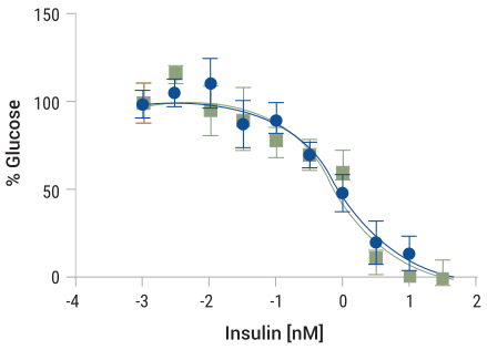
Glucose-Glo™ Assay
グルコースを高感度に測定する生物発光アッセイです。1タイムポイントあたり馴化細胞培養培地2〜5μlのみで測定可能なため、タイムコース試験において同一ウェルから複数回のサンプリングが行えます。マイクロスフェロイド培養の馴化培地での使用が確認されています。
iCell®肝細胞スフェロイドにおけるインスリンを介した糖新生阻害
GSH/GSSG-Glo™ Assay
溶解アッセイ
高感度な生物発光アッセイで、GSH/GSSG比の変化をモニタリングします。並行ウェルでGSH+GSSG総量とGSSGのみをそれぞれ処理し、GSH/GSSG比を算出します。単層培養(5分間振とう)から3D培養(30分間振とう)へは、プロトコルをわずかに変更しますCellTox™ Greenを前段階で使用して、細胞毒性をモニタリングできます。
HCT116スフェロイドの並行ウェルにおける総グルタチオンと細胞生存性のモニタリング
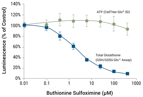
スフェロイドをブチオニンスルホキシミンで48時間処理した後、GSH/GSSG-Glo™ Assayによる総グルタチオン測定またはCellTiter-Glo® 3D Assayによる測定を行った。詳細はサイエンティフィックポスター(下記リンク)をご参照ください。
ROS-Glo™ H2O2 Assay
非溶解アッセイ(オプション)
生物発光 H2O2アッセイにより、活性酸素種をモニタリングします。プロルシフェリン基質が H2O2 と直接反応し、アッセイ成分が活性化されたプロ基質をルシフェリンに変換して生物発光シグナルを生成します。単層培養と3D培養で同じプロトコルが使用可能です。残った細胞は、測定後の細胞は他のセルベースアッセイや核酸抽出に使用できます。
ROS 誘導性のメナジオンに対する HepG2 スフェロイドの応答
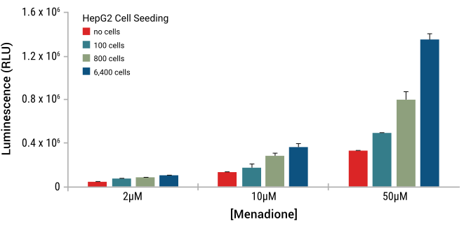
直径の異なるHepG2スフェロイドをさまざまな濃度のメナジオンに暴露したときの応答を示す。細胞は超低接着プレート(Corning)で4日間培養した。H2O2レベルはROS-Glo™ H2O2 Assayで測定し、GloMax® Discoverで読み取った。詳細はアプリケーションノート(下記リンク)をご参照ください。
NAD/NADH-Glo™ およびNADP/ NADPH-Glo™ Assays
溶解アッセイ
この溶解生物発光アッセイで、NAD⁺/NADH比またはNADP/NADPH比をモニタリングします。ライセートを分け、酸/塩基反応によりNAD⁺またはNADHの酸化型もしくは還元型をそれぞれ測定します。標準の単層培養プロトコルからわずかな変更が必要です(詳細は下記ポスターリンクをご参照ください)。
ヒト肝臓マイクロティッシュにおけるトリグリセリド量
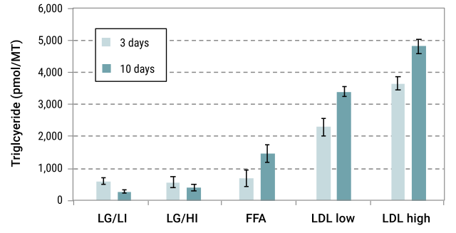
詳細は下記の技術記事をご参照ください。
Lactate-Glo™ Assay
非溶解アッセイ(オプション)
非溶解型の高感度生物発光アッセイにより、3D培養における乳酸の変化をモニタリングします。1タイムポイントあたり培地2〜5μlのみで測定可能なため、同一ウェルから複数回のサンプリングが行えます。測定後の細胞は他のセルベースアッセイや核酸単離に使用できます。
Glutamate-Glo™ Assay
非溶解アッセイ(オプション)
非溶解型の高感度生物発光アッセイにより、3D培養におけるグルタミン酸の変化をモニタリングします。アッセイには、タイムポイントごとに 2〜5μl の培地しか使用しないので、同じウェルから複数回サンプリングすることが可能です。残った細胞は、他の細胞ベースのアッセイまたは核酸単離に使用することができます。
Glutamine/Glutamate-Glo™ Assay
非溶解アッセイ(オプション)
非溶解型の高感度生物発光アッセイにより、3D培養におけるグルタミンおよびグルタミン酸の変化をモニタリングします。グルタミンの測定は、グルタミンからグルタメートへの変換に基づいています。1タイムポイントあたり培地2〜5μlのみで測定可能なため、同一ウェルから複数回のサンプリングが行えます。測定後の細胞は他のセルベースアッセイや核酸抽出に使用できます。
Glycerol-Glo™ Assay
非溶解アッセイ(オプション)
Triglyceride-Glo™ Assay
溶解アッセイ
Cholesterol/Cholesterol Ester-Glo™ Assay
溶解アッセイ
Cholesterol/Cholesterol Ester-Glo™ Assayは、培養細胞ライセートや細胞培養培地などの生体サンプル中のコレステロールおよびコレステロールエステルを迅速かつ高感度に測定するための生物発光アッセイです。コレステロールはステロイド産生、胆汁酸合成、細胞シグナル伝達、膜構造の維持に関わる必須脂質です。
3D細胞培養における生細胞タンパク質ダイナミクスの解析方法
蛍光イメージングとタンパク質間相互作用アッセイにより、3D細胞培養においてタンパク質ダイナミクスを直接観察できます。HaloTag®リガンドとNanoBRET®テクノロジーは、複雑なシステムにおける非破壊的な測定を可能にし、新しいアプローチ方法論(NAMs)に関する規制要件への対応を支援します。 3D細胞培養モデルは、栄養素の勾配、マトリックスとの相互作用、細胞間接触を保持することで、2Dシステムよりもin vivoのタンパク質挙動をより正確に反映します。ただし、こうした複雑さに対応するためには、生きた3次元環境において安定して機能する専用の検出方法が必要です。 規制当局が動物実験の代替手法をますます重視する中、新しいアプローチ方法論(NAMs)はGLP準拠で3Dモデルに対応したアッセイを必要とします。この動向は、創薬開発と毒性スクリーニングにとって重要な変化です。
NanoBRET® Nano-Glo® Detection Systems
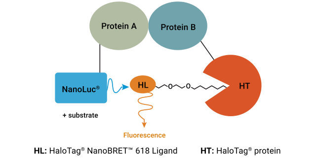
NanoBRET® Assayは、生細胞における2つの結合パートナーの相互作用を測定する、生物発光共鳴エネルギー移動(BRET)ベースのアッセイです。NanoBRET®テクノロジーは、自然な細胞環境におけるタンパク質間相互作用の高感度かつ再現性の高い検出を可能にします。3Dスフェロイドにおけるタンパク質間相互作用の研究へのこのアッセイの応用については、査読済み論文「生細胞における内在性タンパク質間相互作用を定量する生物発光ベースのアッセイ」をご参照ください。
NanoBRET® PPI Assayの原理
3D細胞培養における遺伝子発現の解析方法
3D培養で増殖した細胞は、単層培養と比較して遺伝子発現に差異が生じる場合があります。細胞間接触および細胞-マトリックス間接触の変化は大きく異なり、培養内の酸素と栄養素の勾配もまた遺伝子発現に影響します。遺伝子発現の差異を解析するには、まずRNAを単離・定量する必要があります。その後、特定の遺伝子をRT-qPCRで解析するか、RNA-seqなどの手法で細胞全体の変化をモニタリングします。
注目製品
ReliaPrep™ RNA Miniprep Systems
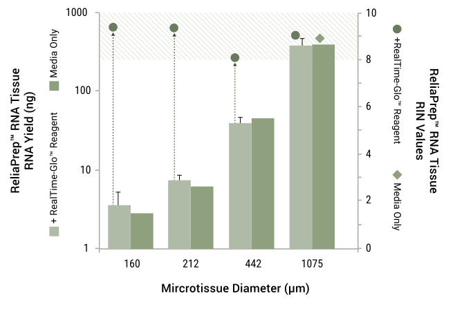
アプリケーションにすぐ使用できる総RNAまたはmiRNAを3D培養から単離し、発現変化をモニタリングします。ReliaPrep™ RNA Miniprep Systemsは、RealTime-Glo™ MT Cell Viability Assayなどの非溶解アッセイとの相乗効果を発揮します。
HEK293マイクロティッシュスフェロイドからのRNA抽出
3D細胞培養におけるゲノム解析の方法
遺伝子型-表現型、または表現型-遺伝子型の違いを理解することは、特に初代細胞や腫瘍を研究する際に重要です。遺伝子型を解析して3D細胞培養で表現型を理解するアプローチもあれば、表現型を研究した後に遺伝子型を調べてその変化の原因を解明するアプローチもあります。ゲノムDNA解析では、まずDNAを抽出・定量します。次に、qPCRで特定の遺伝子を解析するか、NGS(次世代シーケンシング)でゲノム全体を解析します。初代細胞を使用する場合は、各サンプルがどのドナー由来かを識別できることが必須です。そのためSTRプロファイルが広く用いられます。STRプロファイルは、使用している細胞が目的の細胞であることを確認するためにも重要です。
注目製品
Maxwell® RSC Cultured Cells DNA Kit
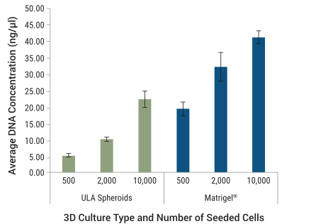
Maxwell® RSCを使用したシンプルなウォークアウェイ型の自動化プロトコルで、3D培養からゲノム解析にすぐ使用できるゲノムDNAを単離します。精製されたDNAは約45分で解析可能な状態になります。単離されたゲノムDNA(gDNA)は、遺伝子コピー数やバイオマーカーのqPCRベース解析、サンプルトラッキングや同一性確認のためのSTRベース解析、およびシーケンシングベース解析に使用できます。
様々な細胞数の HCT116細胞からMaxwell® RSC Cultured Cells DNA Kitで単離した DNAの濃度
3D細胞培養におけるゲノム解析を補完するアッセイ
アッセイ選択マトリックス:3D培養アプリケーション
3Dモデルの種類、ワークフローの要件、下流のアプリケーションに基づいて最適なアッセイを選択するためのマトリックスです。
| アッセイ名 | アッセイ種別 | 溶解/非溶解 | 適した3Dモデル | 下流オプション | マルチプレックス対応 |
|---|---|---|---|---|---|
| CellTiter-Glo® 3D | 細胞生存性(ATP) | 溶解 | スフェロイド、オルガノイドなど | なし | 対応(LDH-Glo™、CellTiter™との併用) |
| RealTime-Glo™ | 細胞生存性 | 非溶解 | すべての3Dモデル | RNA/DNA抽出 | 対応(CellTiter™ Greenとの併用) |
| LDH-Glo™ | 細胞毒性(培地) | 非溶解 | すべての3Dモデル | 細胞生存性測定(後続) | 対応(複数タイムポイント) |
| CellTiter™ Green | 細胞毒性(生細胞) | 非溶解 | すべての3Dモデル | 細胞生存性測定(後続) | 対応(RealTime-Glo™との併用) |
| Caspase-Glo® 3/7 | アポトーシス | 溶解 | スフェロイド、オルガノイド | なし | 対応(CellTiter™、CellTiter-Glo®との併用) |
| Annexin V | アポトーシス+ネクローシス | 非溶解 | すべての3Dモデル | RNA/DNA抽出 | 対応(Caspase-Glo®またはRealTime-Glo™) |
| P450-Glo™ | 薬物代謝 | 非溶解 | 肝細胞スフェロイド | 細胞生存性測定(後続) | 対応(同一プロトコル、併用可能) |
| LC3 HIBIT | オートファジー | 非溶解 | 遺伝子導入済み3Dスフェロイド | 顕微鏡解析 | 細胞生存率アッセイと併用可能 |
| Extracellular ATP | 免疫原性細胞死 | 非溶解 | すべての3Dモデル | 他の細胞死マーカー | 対応(キネティックモニタリング) |
| Glucose-Glo™ | グルコース代謝 | 非溶解 | すべての3Dモデル | 他の代謝アッセイ | 対応(複数タイムポイント/ウェル) |
| NAD/NADH-Glo™ | 酸化還元状態 | 溶解 | スフェロイド、オルガノイド | なし | 並行ウェル(同一ウェルでは不可) |
| Lactate-Glo™ | ラクテート代謝 | 非溶解 | すべての3Dモデル | 他の代謝アッセイ | 対応(1ウェルから複数サンプル) |
| GSH/GSSG-Glo™ | 酸化ストレス | 溶解 | スフェロイド、オルガノイド | なし | 並行ウェル(同一ウェルでは不可) |
| ROS-Glo™ H2O2 | 酸化ストレス | 非溶解 | すべての3Dモデル | 他のストレスマーカー | 対応(下流アッセイ) |
| Glycerol-Glo™ | 脂質代謝 | 非溶解 | 肝細胞、脂肪細胞モデル | 他の脂質アッセイ | 対応 |
| Triglyceride-Glo™ | 脂質蓄積 | 溶解 | 肝細胞、脂肪細胞モデル | なし | 並行ウェル |
| Cholesterol-Glo™ | 脂質代謝 | 溶解 | 肝細胞、ステロイド産生細胞モデル | なし | 並行ウェル |
FAQ
3D細胞培養に標準のCellTiter-Glo® AssayまたはCellTiter-Glo® 2.0を使用できますか?
CellTiter-Glo® 3D Cell Viability Assayをご使用ください。このアッセイは3Dモデル向けに特化して最適化されており、完全な溶解を保証します。CellTiter-Glo®またはCellTiter-Glo® 2.0は、3Dモデルの完全溶解に十分な溶解能を持たない場合があり、アッセイ結果に意図しないバイアスやばらつきの増大をもたらす可能性があります。
スフェロイドとオルガノイドでは、どのアッセイを使用すべきですか?
スフェロイドとオルガノイドはいずれも、プロメガの3Dアッセイの大半に対応しています。主な違いは次の通りです:スフェロイドはよりシンプルで、一般的に単一の細胞種で構成されます;オルガノイドは複数の細胞種を含み、固有の組織構造を持ちます。オルガノイドでは、組織構造を保持するために非溶解アッセイ(RealTime-Glo™ MT、代謝活性アッセイ)がより適している場合が多いです。最適化されたプロトコルについては、各アッセイのアプリケーションノートをご参照ください。
同一の3D培養ウェルでアッセイをマルチプレックス解析できますか?
アッセイによって異なりますが、同一ウェルでのマルチプレックス解析は複数の3Dアッセイにおける大きな利点です。例えば、RealTime-Glo™ MTで細胞生存性を測定した後、同一ウェルにCellTox™ Greenを添加して死細胞を検出し、キネティック測定を継続することができます。また、同一ウェルからDNAまたはRNAを抽出して、ゲノム特性解析や発現解析に活用することも可能です(例:ReliaPrep™ Miniprep systemsやMaxwell® RSCキットの使用)。アッセイの適合性は必ずテクニカルマニュアルで確認するか、プロメガ テクニカルサービスにお問い合わせください。
細胞生存性の測定にはスフェロイドを溶解する必要がありますか?
必ずしも必要ではありません。ワークフローに基づいて選択してください。溶解アッセイ(CellTiter-Glo® 3D, NAD/NADH-Glo™)はスフェロイドを完全に溶解してコアまで浸透しますが、その後のアッセイ実施はできなくなります。非溶解アッセイ(RealTime-Glo™ MT、代謝アッセイ)は細胞を保存するため、キネティック測定や複数回測定のワークフローに最適です。また、非溶解アッセイでは他のアッセイとのマルチプレックス解析も可能で、スフェロイドの生物学的な側面(代謝マーカーなど)を多角的に評価したり、下流のDNA/RNA抽出を行うこともできます。
3DでのHTS(ハイスループットスクリーニング)に対して検証済みのアッセイはどれですか?
プロメガは、ハイスループットに対応するよう特化して設計された生物発光アッセイの豊富なラインナップを提供しています。CellTiter-Glo® 3D、Caspase-Glo® 3/7 3D Assayなどがその代表例です。これらのアッセイはHTSに対応し、384ウェルプレートへのスケールアップも可能です。複数用量の試験は並行ウェルで実施できます。推奨タイムポイント、プレートの種類、3D専用の最適化のためのGloMax®プレートリーダー設定については、アプリケーションノートをご参照ください。
同一の3D培養ウェルを複数のアッセイ測定に使用できますか?
非溶解アッセイを使用すれば可能です。ワークフロー例:(1)Day 0〜3:RealTime-Glo™ MTによるキネティック細胞生存性モニタリング;(2)Day 3:培地上清のサンプリングによる代謝活性スクリーニング(例:別プレートで培地5μlからグルコースおよび/またはラクテートを測定);(3)Day 5:無傷のスフェロイドからRNAを抽出;(4)Day 5:抽出したRNAでRT-qPCRを実施。溶解アッセイはサンプルを消費するため、下流の解析は行えません。
3D培養アッセイの一般的な測定期間はどのくらいですか?
スフェロイド/オルガノイドは一般的に、播種後48時間〜7日の間に測定します。キネティックアッセイ(RealTime-Glo™ MT)はこの期間を通じて継続的なモニタリングが可能です。溶解アッセイ (CellTiter-Glo® 3D)はエンドポイント測定(3〜7日)に最適です。細胞の種類やモデルによって最適化が必要な場合があります。具体的な推奨事項については、各アッセイのアプリケーションノートをご参照ください。
プロメガの3Dアッセイはさまざまなプレートタイプに対応していますか?
はい。超低接着プレート(ULA)はスフェロイド培養の標準であり、すべてのアッセイに対応しています。ハンギングドロッププレートも使用可能です。オルガノイドについては、培養方法によって選択が異なります。ハイドロゲルベースの培養は対応していますが、アッセイ固有のテクニカルドキュメントで確認してください。プレートの形状は浸透のキネティクスに影響する場合があります。プレートの適合性を確認するため、必ずご使用のモデルシステムでテストを実施してください。
プロメガの3Dアッセイはどのプラットフォームで測定できますか?
GloMax®リーダーは発光アッセイの主要プラットフォームです。GloMax® Discoverは、マルチプレックス解析向けに発光と蛍光の組み合わせ測定に対応しています。72時間を超えるキネティック測定では、適切な環境条件下で細胞が培養されていることを確認してください。蛍光アッセイ(例:CellTox™ Green)は、あらゆる蛍光プレートリーダーで測定可能です。
スフェロイドやオルガノイドなどの3D培養システムでゲノム解析を行うには?
3D培養のゲノム解析は、回収、核酸抽出、下流解析の3つの主要ステップで行います
- ステップ1. 細胞をマトリックスから回収します(例:ハイドロゲルの溶解、必要に応じて酵素処理によるスフェロイドの解離)。スフェロイドやオルガノイドの細胞外マトリックスと密な構造は収量を低下させ、阻害因子を捕捉することがあるため、穏やかかつ十分な溶解が重要です。
- ステップ2. DNAまたはRNAを抽出します。インプット量が少ない貴重なサンプルには、Maxwell®装置・キットなどの自動化システムが反復間の再現性を向上させます。ReliaPrep™ Miniprepシステムなどのカラムベースの選択肢は、コスト効率の高い手動ワークフローや少数サンプルに適しています。3D構造体は2D単層培養よりも得られる材料が少ない場合が多いため、複数のオルガノイドをプールするか、少量インプット用に検証されたキットの選択を検討してください。
- ステップ3. 解析を実施します。抽出した核酸は、遺伝子発現・変異・クローン不均一性の解析のために、qPCR、RT-qPCR、全ゲノムシーケンシングまたはRNAシーケンシング、メチル化解析、遺伝子型解析に供することができます。
限られたサンプルから最大限のデータを得るために、上流の細胞生存性アッセイや細胞毒性アッセイと同一のウェルからDNA/RNAを抽出することも可能です。事前に必ず適合性を確認してください。使用するモデルや下流アプリケーションに適した抽出方法については、プロメガ テクニカルサービスにお問い合わせください。
3D培養における生細胞タンパク質ダイナミクスはどのように測定できますか?
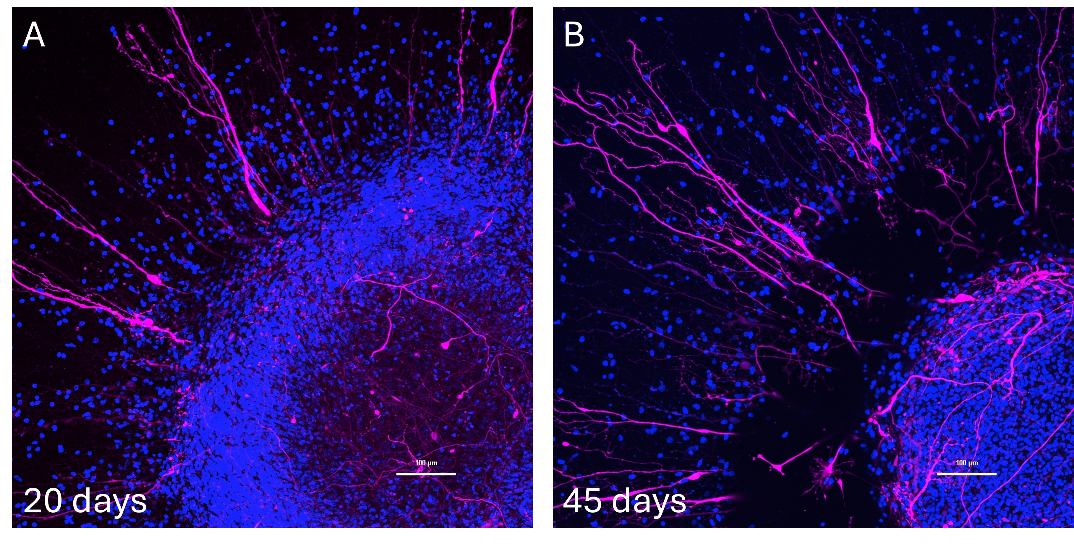
HaloTag®およびNanoLuc®レポーターを使用することで、モデルを破壊することなく、生きたスフェロイドやオルガノイドにおけるタンパク質の存在量、局在、ターンオーバーを追跡できます。HaloTag®は細胞透過性のJanelia Fluor®リガンドと組み合わせることで、先進的な3Dモデルにおけるタンパク質の局在・ターンオーバーの生細胞イメージング(パルスチェイス研究を含む)を可能にします。NanoLuc®レポーターとの組み合わせにより、エンドポイントアッセイでは対応が難しい3Dシステムのリアルタイム非破壊モニタリングを実現します。プロメガの研究者たちは、HaloTag®-NanoLuc®融合体とJanelia Fluor®リガンドを使用して、数日間のタイムコースにわたって脳スフェロイドにおけるタンパク質発現の定量とリアルタイムの神経突起形成測定を実施しました。リアルタイムの3Dイメージングでは、スフェロイドの生存性を保ちながら数日間持続するシグナルが必要なため、ご使用のモデルでリガンドと基質の安定性および浸透性を確認してください(Q1参照)。
3D培養に特化したガイダンスについては、ホワイトペーパー「NanoLuc®とHaloTag®テクノロジーを使用した脳オルガノイドのリアルタイムモニタリング」をご参照ください。
生理学的により関連性の高い研究ツールをお探しですか?
タンパク質ダイナミクスと細胞活性を検出するためのツールの全ラインナップをご覧ください。