Caspase-Glo® 1 Inflammasome Assay
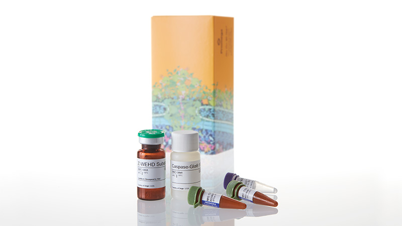
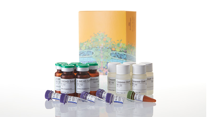
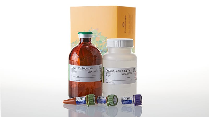
A Bioluminescent Method to Directly Measure Inflammasome Activity
- Fast, simple, homogeneous caspase-1 assay kit
- Selective and quantitative
- Multiplex and high-throughput screening (HTS) compatible

Catalog Number:
Size
Catalog Number: G9951
Catalog Number: G9952
Catalog Number: G9953
Caspase-Glo® 1 の原理は?
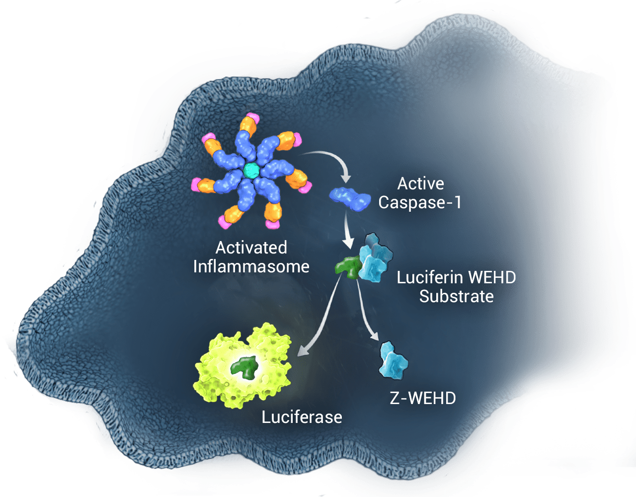
Caspase-Glo® 1 Inflammasome Assay はインフラマソームの必須構成要素でカスパーゼのメンバーであるカスパーゼ-1 の活性を選択的に測定するシンプルでホモジニアスな発光法です。インフラマソームは多様な炎症性刺激により誘導されるタンパク質複合体です。自然免疫細胞は病原体やその他の危険信号に応答してインフラマソームを形成し、プロカスパーゼ-1前駆体が活性化カスパーゼ-1に変換されます。
カスパーゼ -1 活性化の結果、サイトカイン IL-1β および IL-18 のプロセシングと放出が起こり、免疫原性の細胞死であるパイロトーシスとなります。 Caspase-Glo 1 assay は細胞内や培地中のカスパーゼ -1 活性を直接測定できる感度を有しています。
革新的なケミストリ
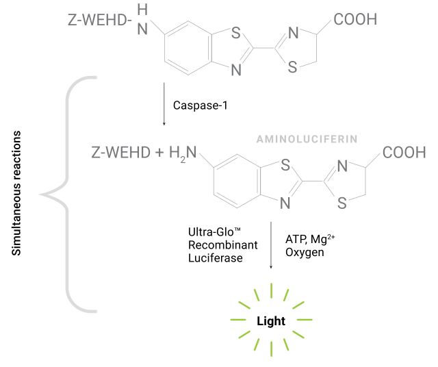
カスパーゼによる Z-WEHD 基質 (Z-WEHD-アミノルシフェリン) の切断によりルシフェラーゼの基質(アミノルシフェリン)が放出され、特別な耐熱性組換えルシフェラーゼにより光が生成されます。
Fast and Simple
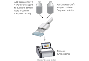
No sample preparation or manipulation required. Simply add, mix and then measure luminescence after only one hour. You can get results faster in fewer steps compared to conventional Western blot and ELISA analyses. The stable luminescent signal and large volume (100ml) format supports high-throughput screening applications.
Selective and Quantitative
The selective caspase-1 substrate (Z-WEHD) enables detection of catalytically active caspase-1 in cells or culture media and quantitative measurement of inflammasome activity. Inclusion of the proteasome inhibitor MG-132 in the reagent eliminates non-specific proteasome substrate cleavage, enabling sensitive detection of caspase-1. The assay includes a caspase-1 specific inhibitor (Ac-YVAD-CHO) to confirm specific activity in parallel samples and distinguish caspase-1 activity from other caspases.
Caspase-1 Activity is Distinguished From Other Caspases
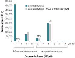
Inflammation and apoptosis caspases at equimolar concentrations were tested with the assay, +/– Ac-YVAD-CHO, and caspase activity was measured after 20 minutes. Ac-YVAD-CHO at 1µM inhibited 99% of the caspase-1 activity, whereas it had minimal effect on the other cross-reacting caspases (5, 3 and 6).
NLRP3 Inhibitor Prevents Caspase-1 Activation in THP-1 and PBMC Cells
NLRP3 plays a key role in immune sensing by initiating the assembly of an inflammasome in response to various danger signals. The NLRP3 inflammasome triggers caspase-1 activation and IL-1β cytokine secretion, eventually resulting in an inflammatory, pyroptotic cell death. The Caspase-Glo® 1 Assay allows you to easily measure the effect of NLRP3 inhibitors on caspase-1 activation in primary cells.
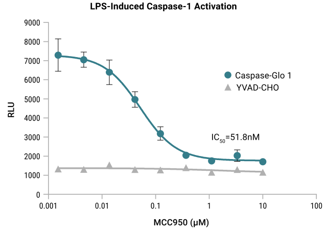
THP-1 cells were differentiated for 3 days with PMA. Medium was replaced and MCC950 (NLRP3 inflammasome inhibitor), was added followed by LPS or vehicle. Half of the culture medium (50µl) was removed after 3 hours of LPS and tested with Caspase-Glo® 1 Reagent or Caspase-Glo® 1 Reagent + YVAD-CHO.
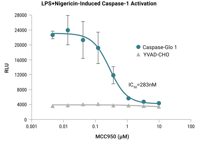
PBMCs were plated, then treated with MCC950 followed by LPS or vehicle overnight. Following the overnight treatment, cells were treated with nigericin or vehicle. Nigericin causes potassium efflux and significant caspase-1 activation after LPS priming. Half of the culture medium (50µl) was then removed and tested with Caspase-Glo® 1 Reagent or Caspase-Glo® 1 Reagent + YVAD-CHO. Luminescence was read for both after 1 hour. Caspase-Glo® 1 signal demonstrates a dose-dependent inhibition of caspase-1 activity by the NLRP3 inflammasome inhibitor.
Caspase-1 Activity Detected in Peripheral Blood Mononuclear Cells and Released into Medium
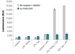
Caspase-Glo® 1 Inflammasome Assay monitors caspase-1 activity in peripheral blood mononuclear cell (PBMC) supernatant. Cells were stimulated with or without LPS then subsequently stimulated with ATP for 30 minutes. Cells were then pelleted and supernatant was assayed for caspase-1 activity.
Multiplex With Other Cell-Based Assays
The Caspase-Glo® 1 Assay is sensitive enough to detect caspase-1 activity from a small volume of transferred medium. This leaves cells intact for other assays, such as cell viability or cytotoxicity, so you can get more data per sample.
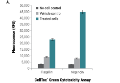
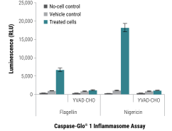
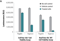
Caspase-Glo® 1 Inflammasome Assay Multiplexed with Cell Viability and Cell Death Assays. THP-1 cells were grown in RPMI 1640 medium supplemented with 10% FBS in a 37°C incubator with 5% CO2. Cells were added to plates at 5 x 10⁵ cells/ml in 100µl of medium and differentiated with PMA (20nM, 3 days) in 96-well plates followed by treatment with flagellin (1µg/ml, 1 hour) or nigericin (20µM, 2 hours). In Panel A CellTox™ Green Reagent, Caspase-Glo® 1 Reagent or Caspase-Glo® 1 YVAD-CHO Reagent was added to the cells, and fluorescence (CellTox™ Green) or luminescence (Caspase-Glo® 1) was recorded after 90 minutes. In Panel B cell viability was monitored with CellTiter-Glo® or RealTime-Glo™ MT Cell Viability Assay. Luminescence was recorded at 10 minutes and 90 minutes, respectively.
Featured Publications
This peer-reviewed paper "A bioluminescent caspase-1 activity assay rapidly monitors inflammasome activation in cells" shows how the Caspase-Glo® 1 Assay can specifically detect caspase-1 activity in cells treated with inflammasome inducers, confirm one-signal and two-signal inflammasome activation mechanisms, and monitor caspase-1 activity, cell death, and IL-1β release from the same microplate well.
This peer-reviewed study "Interrogating direct NLRP3 engagement and functional inflammasome inhibition using cellular assays" examines the direct engagement and functionality of NLRP3 pathway activity in cells using several mechanistic assays, including the Lumit® IL-1β Immunoassay and Caspase-Glo® 1 Inflammasome Assay.
Protocols
Complete Protocol
Specifications
Catalog Number:
選択製品の構成品内容
| Item | Part # | Size | Concentration |
|---|---|---|---|
|
MG-132 Inhibitor |
G932B | 2 × 30μl | |
Z-WEHD Substrate |
G991A | 1 × 1 bottle | |
Ac-YVAD-CHO |
G992A | 1 × 10μl | 2mM |
Caspase-Glo® 1 Buffer |
G994A | 1 × 10ml |
選択製品の構成品内容
| Item | Part # | Size | Concentration |
|---|---|---|---|
|
MG-132 Inhibitor |
G932C | 1 × 300μl | |
Z-WEHD Substrate |
G991A | 5 × 1 bottle | |
Ac-YVAD-CHO |
G992A | 5 × 10μl | 2mM |
Caspase-Glo® 1 Buffer |
G994A | 5 × 10ml |
選択製品の構成品内容
| Item | Part # | Size | Concentration |
|---|---|---|---|
Caspase-Glo® 1 Buffer |
G994B | 1 × 100ml | |
|
MG-132 Inhibitor |
G932C | 2 × 300μl | |
Z-WEHD Substrate |
G991B | 1 × 1 bottle | |
Ac-YVAD-CHO |
G992B | 1 × 100μl | 2mM |
Resources
Related Products
類似製品
Lumit® IL-1β Human/Mouse Immunoassay
シンプルな無洗浄プロトコルを使用して、細胞培養サンプル中に放出されたIL-1βを定量的に測定。
W6010, W6011, W6012, W7010, W7011, W7012, W116A-C, W119A-C
Lumit® Active IL-18 (Human) Immunoassay
シンプルで洗浄不要のプロトコルを使用して、70分以内に細胞培養サンプル中の活性化IL-18を定量。
W1910, W1911, W1912, W191A-C
CellTox™ Green Cytotoxicity Assay
細胞死に伴う細胞膜破壊により細胞毒性を蛍光で測定
G8741, G8742, G8743, G8731
CellTiter-Glo® 2.0 Cell Viability Assay
試薬の安定性が向上したCellTiter-Glo®、ATP検出に基づく細胞増殖の定量が可能
G9241, G9242, G9243
関連製品
GloMax® Discover System
発光・蛍光・吸光度測定に対応した高性能マイクロプレートリーダー
GM3000
Caspase-Glo® 3/7 Assay System
カスパーゼ-3/7活性の検出に対応した、使いやすいプレートベースの発光アッセイ
G8090, G8091, G8093, G8092
LDH-Glo™ Cytotoxicity Assay
3Dマイクロティッシュなど細胞数の少ないサンプルに対応した、高感度な発光ベースLDH細胞傷害性アッセイ
J2380, J2381
Lumit® TNF-α (Human) Immunoassay
Quantitatively measures released TNF-α in cell culture samples using a simple, no-wash protocol.
W6050, W6052, W6051, W137A-C