ONE-Glo™ + Tox Luciferase Reporter and Cell Viability Assay
Sensitive Detection of Reporter Activity and Cell Health
- Measures cell viability and firefly luciferase activity in the same assay well
- Easy to use—simply add reagent, mix and read
- Scalable to meet throughput needs, up to 1,536-well format

Catalog Number:
Size
Catalog Number: E7110
Catalog Number: E7120
Understand Reporter Gene Activity in the Context of Cell Viability
アッセイの第一段階では、実験処理後の培養細胞群における生細胞の相対数を非溶解性の蛍光アッセイ(CellTiter-Fluor™ Cell Viability Assay)で測定します。CellTiter-Fluor™ Assayは、生細胞内に常在するプロテアーゼ活性を細胞生存性マーカーとして測定します。細胞透過性の蛍光基質 (glycylphenylalanyl-aminofluorocoumarin; GF-AFC) がインタクトな細胞に入り込むと生細胞プロテアーゼにより切断され、生細胞数に比例した蛍光シグナルを生成します。この生細胞プロテアーゼは細胞膜の完全性が失われ培地中に漏出すると不活性化されます。遊離したAFC蛍光物質の蛍光はマイクロプレートリーダーやCCDイメージャー(励起波長:380–400nm、蛍光波長:505nm) で測定します。
アッセイの第二段階では、発現したホタルルシフェラーゼ酵素の測定にONE-Glo™ Luciferase Assay Systemを用います。
このシステムに含まれるONE-Glo™ Luciferase Assay Buffer および ONE-Glo™ Luciferase Assay Substrateを混和して ONE-Glo™ Reagentを調製します。 ハイスループットあるいはウルトラハイスループットなアプリケーションに対応するために新規なfluoroluciferin substrateが採用されており、試薬の安定性が向上し、サンプルに含まれる成分に対する耐性が得られます。また、標準的なルシフェラーゼアッセイ試薬よりも硫黄臭が抑えられています。発光はマイクロプレートリーダーやCCDイメージャーで測定します。
また、恒常的な発現プロモーターを有するホタルルシフェラーゼレポーターベクターを使用することでタンパク質の発現効率と細胞生存性を加味したトランスフェクション条件の最適化にも利用することができます。
- 効率的:細胞生存性とルシフェラーゼ遺伝子発現を同一アッセイウェル内で測定。単一ウェル内の細胞で複数項目のデータ取得。
- より生物学的な情報を取得:細胞生存性を背景としたレポーター遺伝子発現の理解
- 簡便:アッセイはシンプルで連続的な“添加-混和-測定”フォーマットを採用
- 高い柔軟性と容易な自動化:各アッセイ試薬の容量はサンプル処理量に合わせてスケールを変更でき、最大1536ウェルフォーマットまで自動化可能
- Niles, A.L. et al. (2007) A homogeneous assay to measure live and dead cells in the same sample by detecting different protease markers. Anal. Biochem. 366, 197–206.
Step 1: Cell Viability Assay
In the first part of the assay, a nonlytic fluorescence-based method (CellTiter-Fluor™ Cell Viability Assay) is used to measure the relative number of live cells in culture. The CellTiter-Fluor™ Assay measures a conserved and constitutive protease activity within living cells. This live-cell protease activity is restricted to intact viable cells and is measured using a fluorogenic, cell-permeant peptide substrate (glycylphenylalanyl-aminofluorocoumarin; GF-AFC). The substrate enters intact cells where it is cleaved by the live-cell protease to generate a fluorescent signal proportional to the number of living cells. The live-cell protease becomes inactive upon loss of cell membrane integrity and leakage into the surrounding culture medium. Fluorescence of the free AFC fluorophore is measured with a microplate reader or CCD imager using an excitation wavelength of 380–400nm and emission wavelength of 505nm.
Step 2: ONE-Glo™ Luciferase Assay
The second part of the assay uses the ONE-Glo™ Luciferase Assay System to quantify firefly luciferase gene expression from cells expressing the reporter. Ideally suited for high- and ultrahigh-throughput applications, the ONE-Glo™ Assay Reagent contains a fluoroluciferin substrate that gives increased stability and greater tolerance of sample components, and has less odor than standard luciferase assay reagents. Luminescence is measured with a microplate reader or CCD imager.
Reference
Niles, A.L. et al. (2007) A homogeneous assay to measure live and dead cells in the same sample by detecting different protease markers. Anal. Biochem. 366, 197–206.
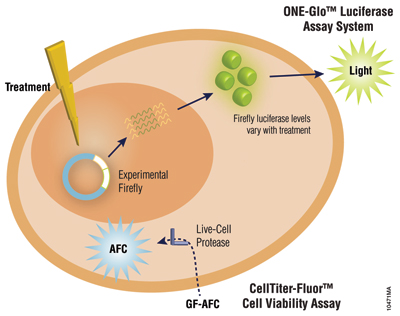
Schematic of the ONE-Glo™+Tox Luciferase Reporter and Cell Viability Assay.
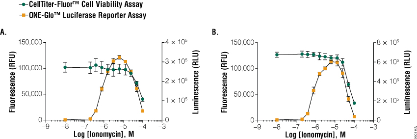
Example ONE-Glo™ + Tox Assay Data
Ionomycin Titration in 96- and 384-well Plates
In the experiment shown here, 1 × 10⁴ (96-well plate; Panel A) or 5 × 10³ (384-well plate; Panel B) cells expressing NFAT response element were treated with serial titrations of ionomycin in the presence of PMA for 6 hours. At specific concentrations, ionomycin and PMA work cooperatively to stimulate NFAT-dependent gene expression. However, higher concentrations of ionomycin result in cytotoxicity seen as a decrease in viability (fluorescence). A decrease in reporter expression (luciferase) activity is also observed due to the increase in cytotoxicity.
Specifications
Catalog Number:
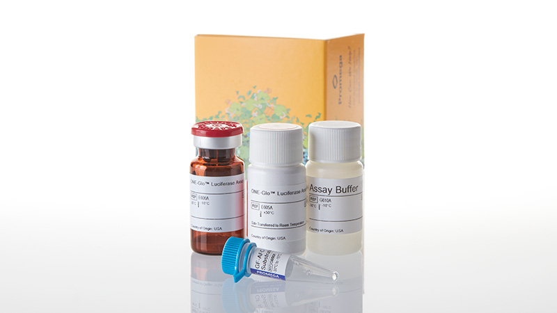
選択製品の構成品内容
| Item | Part # | Size |
|---|---|---|
|
ONE-Glo™ Luciferase Assay Buffer |
E605A | 1 × 10ml |
|
ONE-Glo™ Luciferase Assay Substrate |
E606A | 1 × 1 vial |
|
GF-AFC Substrate |
G608A | 1 × 10μl |
|
Assay Buffer |
G610A | 1 × 10ml |
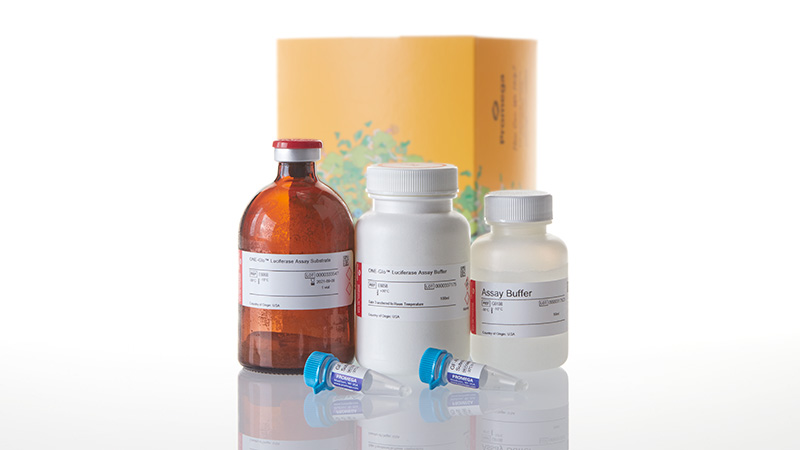
選択製品の構成品内容
| Item | Part # | Size |
|---|---|---|
|
ONE-Glo™ Luciferase Assay Buffer |
E605B | 1 × 100ml |
|
ONE-Glo™ Luciferase Assay Substrate |
E606B | 1 × 1 vial |
|
GF-AFC Substrate |
G608B | 2 × 50μl |
|
Assay Buffer |
G610B | 1 × 50ml |
Resources
No related resources available
Related Products
類似製品
ONE-Glo™ EX Luciferase Assay System
ONE-Glo™ Assayの改良版で高感度・長時間発光、保存方法の選択肢も増えました
E8110, E8120, E8130, E8150
RealTime-Glo™ MT Cell Viability Assay
72 時間まで培養細胞の生存性をモニタリングできる発光アッセイ
G9711, G9712, G9713
ApoTox-Glo™ Triplex Assay
同じサンプルウェルより細胞生存性、細胞毒性、アポトーシスが測定できるトリプルアッセイ
G6320, G6321
Nano-Glo® Dual-Luciferase Reporter Assay System
シングルサンプル中のホタルおよび NanoLuc® ルシフェラーゼ活性を高感度に検出
N1610, N1620, N1630, N1650, N1521, N1531, N1541, N1551
関連製品
Promoterless Firefly Luciferase Basic Vectors
pGL4.10[luc2]、pGL4.11[luc2P]、pGL4.12[luc2CP]ベクターはそれぞれ安定性の異なるルシフェラーゼタンパク質をコードし、遺伝子発現転写調節領域の研究に使用できます。
E6651, E6661, E6671
ViaFect™ Transfection Reagent
iPS細胞など様々な細胞株に使える、高効率・低毒性なトランスフェクション試薬
E4981, E4982
GloMax® Discover System
発光・蛍光・吸光度測定に対応した高性能マイクロプレートリーダー
GM3000
FuGENE® HD Transfection Reagent
トランスフェクション困難な細胞株を含む広範な細胞腫に、より優れたパフォーマンス
E2311, E2312