Anti-LgBiT Monoclonal Antibody
Detects Large BiT (LgBiT) Subunit and LgBiT Fusion Proteins
- Affinity-purified mouse monoclonal antibody
- Use in Western blotting and immunofluorescence
- Detects LgBiT subunit used in NanoBiT® complementation technology

Catalog Number:
Size
Catalog Number: N7100
Detect LgBiT Subunit of NanoBiT® Complementation System
Anti-LgBiT Monoclonal Antibody is a protein A/G affinity-purified mouse monoclonal antibody that is used to detect Large BiT (LgBiT) and LgBiT fusion proteins via Western blotting and immunofluorescence. Weak cross-reactivity with NanoLuc® luciferase is observed.
LgBiT is the large 18kDa subunit of NanoBiT® complementation technology. LgBiT interacts with the low-affinity SmBiT or the high-affinity HiBiT peptides to form a functional luciferase. LgBiT protein fusions are used with SmBiT protein fusions for live cell analysis of protein interactions. Unfused LgBiT can also be expressed in cells to detect HiBiT-tagged proteins without the need for cell lysis.
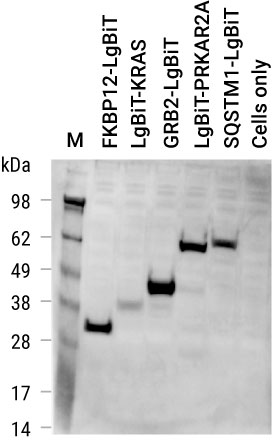
Western Blot Results
The figure on the right shows an example Western blot using HEK293 cells transfected with CMV-based expression constructs encoding LgBiT fusion proteins. The expected mobility was seen for each of the LgBiT fusion proteins. We recommend a concentration of 1µg/ml as a starting point for protocol optimization. Low levels of the LgBiT-KRAS fusion were detected even with CMV-driven expression and without dilution using Transfection Carrier DNA.
HEK293細胞で発現したNanoLuc®融合タンパク質の検出
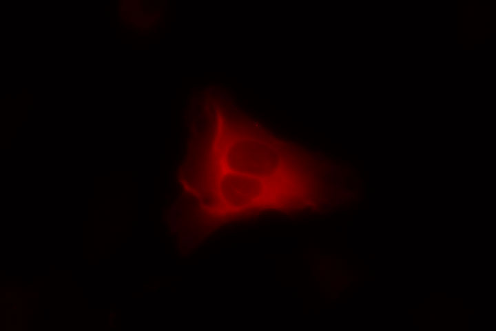
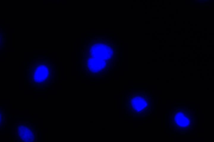
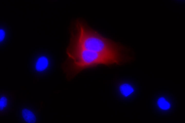
Immunofluorescence Results
Shown below are example immunofluorescent images of HeLa cells transfected with a CMV-based construct expressing LgBiT-CRAF fusion protein. The LgBiT-CRAF fusion protein is stained red, and nuclei are marked blue via Hoechst staining. We recommend a concentration of 1-3µg/ml for immunofluorescence optimization. No LgBiT staining is observed in HeLa cells transfected with only carrier DNA (mock).
LgBiT-CRAF
(3µg/ml Anti-LgBiT)
LgBiT
Hoescht
Merge
Mock (3µg/ml Anti-LgBiT)
LgBiT

Hoescht
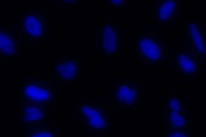
Merge
Protocols
Complete Protocol
Specifications
Catalog Number:
選択製品の構成品内容
| Item | Part # | Size |
|---|---|---|
Anti-LgBiT Monoclonal Antibody |
N710A | 1 × 100μg |
Related Products
関連製品
Nano-Glo® Live Cell Assay
NanoBiT® または NanoLuc® の発光を、生きた細胞で最大2時間測定
N2011, N2012, N2013
Nano-Glo® HiBiT Lytic Detection System
細胞内の HiBiT-タグ融合タンパク質総量を発光法により検出
N3030, N3040, N3050
Nano-Glo® HiBiT Extracellular Detection System
細胞表面や培地中に分泌されたHiBiT タグタンパク質を生細胞のまま発光検出
N2420, N2421, N2422
NanoBiT® PPI Starter Systems
生細胞でタンパク質相互作用の検出を行うための相補的レポーター
N2014, N2015, N2016