Lumit® Insulin Immunoassay
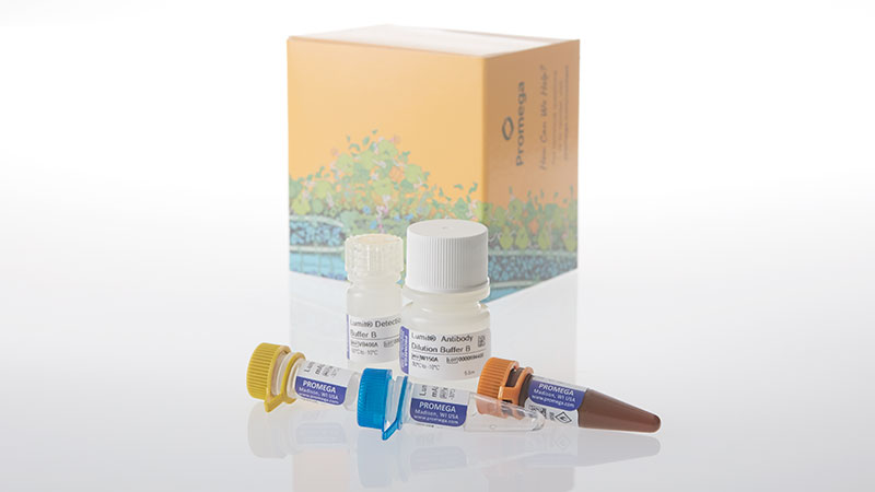
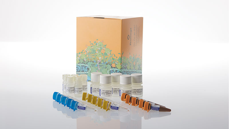
No-Wash Immunoassay for Insulin Detection
- Fast add-and-read protocol
- Luminescent detection with just a plate-reading luminometer
- Flexible, scalable throughput: Perform 100 assays in 96-well format or 400 assays in 384-well format using W8010; perform 500–2000 assays with W8012
Catalog Number:
Size
Catalog Number: W8010
Catalog Number: W8012
Detect Insulin Secretion Without Tedious Wash Steps
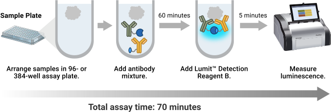
The Lumit® Insulin Immunoassay quantitatively measures insulin from cell culture or islet secretion samples with a fast, easy, no-wash protocol. Simply add Lumit® reagents to the sample, incubate and read using a standard multimode plate reader. Results are available in just 70 minutes. Lumit® assays are ideal for quantitating hormone secretion from islet perifusion workflows. The flexible assay format can be scaled to 384-well plates for high-throughput sample processing.
How the Lumit® Insulin Immunoassay Works
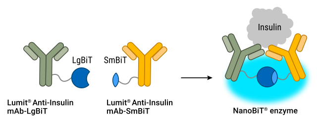
Primary antibodies to insulin, selected for their specificity and sensitivity, are labeled with the LgBiT and SmBiT subunits of NanoBiT® Luciferase. In the presence of insulin, the subunits are brought together to reconstitute active luciferase enzyme. Adding the optimized luciferase substrate generates a bright luminescent signal that is proportional to analyte levels.
Simple One-Plate Protocol, No Wash Steps
Recommended Multiwell Assay Formats
|
Plate Format |
Insulin Sample |
Total Reaction |
Number of Assays |
Number of Assays |
|
96-Well |
50µl |
125µl |
100 |
500 |
|
384-Well |
12.5µl |
31.1µl |
400 |
2000 |
Lumit® assay setup is highly flexible and can be scaled up or down as desired. In addition to the configurations listed above, the assay is compatible with any sample volume ≥5µl provided proportionate amounts of Lumit® reagents are used. For more information, see Table 1 in the Lumit® Insulin Immunoassay Technical Manual (#TM776).
Lumit™ イムノアッセイ vs. ウェスタンブロッティング
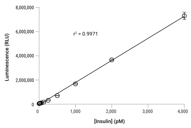
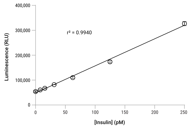
|
Specification |
Lumit® Insulin Immunoassay |
|
|
Limit of Detection (LOD) |
10pM |
|
|
Range |
10pM–8nM |
|
|
Signal/Background max |
>200 |
|
|
Assay Time |
70 minutes |
|
|
Sample Type |
Cell culture supernatants |
|
SD=Standard Deviation
Note: Lumit® Insulin Immunoassay kits do not include a positive control. Instructions for preparing your own positive control for use with the assay can be found in TM776.
Applications for the Lumit® Immunoassays
Insulin and glucagon are key target analytes measured in metabolism research. Secreted from β and α cells of pancreatic islets, respectively, the two hormones are vital for regulating glucose levels in the body. Using the Lumit® Insulin and Lumit® Glucagon Immunoassays, it is easy to collect hormone secretion data from monolayer cells in culture, 3D islet microtissues and islets. The simple add-and-read protocol is compatible with static and dynamic glucose-stimulated insulin secretion (GSIS) experiments, including perifusion systems.
Perifusion is a powerful method for studying hormone secretion over time and in response to sequential treatments, and it generates a large number of samples for analysis. Lumit® Immunoassays make analysis of large sample sets like this much easier by avoiding tedious wash steps. The faster time to result gives you data sooner and enables you to move on to your next experiment.
Quantitate Insulin and Glucagon Secretion from Mouse Pancreatic Islets
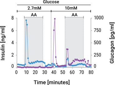
Insulin and glucagon secretion were measured in samples collected during perifusion experiments. Briefly, 80 mouse islets were placed in triplicate chambers of a perifusion instrument (Biorep, Miami Lakes, Florida). The islets were treated with 2.7mM glucose and then 10mM glucose, in combination with an amino acid mixture. Perifusate was collected every minute. Ten microliters of each sample were transferred into wells of a 384-well plate and assayed for either insulin or glucagon. Insulin and glucagon data sets are superimposed. This data was kindly provided by Drs. Hannah Foster and Matthew Merrins, University of Wisconsin VA Hospital, Madison, Wisconsin.
Featured Application Note
Measuring GLP-1 Receptor Agonist Activity in Cells with the Lumit® Insulin Immunoassay
Imperial College London used the Lumit® Insulin Immunoassay to differentiate the potency and activity of three GLP-1 receptor agonist drug candidates, demonstrating that the assay is well-suited for high-throughput metabolic disease drug discovery workflows.
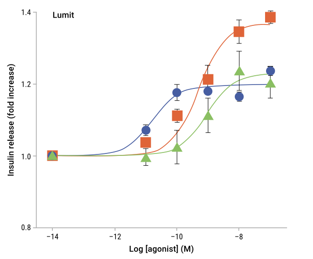
Cells were treated with three GLP-1 receptor agonist drug candidates and insulin secretion measured using the Lumit® Insulin Immunoassay. Each compound demonstrated distinct potency and maximal response, highlighting the assay's ability to discriminate pharmacological differences across candidates.
Protocols
Complete Protocol
Frequently Asked Questions
The complete protocol for this product is available in the Technical Manual (#TM776).
How does the Lumit® Insulin Immunoassay work? How does it differ from a traditional ELISA?
The Lumit® Insulin Immunoassay uses a bioluminescent detection principle rather than enzymatic colorimetric detection. Two antibodies, each labeled with a complementary subunit of NanoLuc® Luciferase, bind to insulin in your sample. When both antibodies bind the same insulin molecule, the subunits reconstitute into an active luciferase enzyme, generating a luminescent signal proportional to insulin concentration. Because reconstitution only happens in the presence of analyte, there are no wash steps required. The total assay time is 70 minutes using a standard multimode plate reader, while ELISA measurement of insulin may take hours.
Should I measure cell viability before measuring insulin secretion?
Yes. In metabolic disease research, viability tells you how to read your functional data. The CellTiter-Fluor™ Cell Viability Assay is non-lytic and doesn't utilize ATP, so you can read viable cell number after compound exposure and then run the secretion assay in the same well. Including a viability assay in your workflow distinguishes a compound that impairs beta cell function from one that simply reduces viable cell number. Read viability after treatment; then move to functional readouts like insulin secretion.
What sample types are compatible with the Lumit® Insulin Immunoassay?
The Lumit® Insulin Immunoassay is validated for cell culture supernatants from monolayer cell culture and islet secretion samples. Sample volumes as low as 5µl can be used if reagent amounts are adjusted proportionately (see Table 1 in TM776 for scaling guidance).
What throughput formats does this assay support?
For optimum performance, use opaque, white multiwell plates compatible with your luminometer. The assay can run in either 96-well or 384-well format:
- 96-well format: 50µl sample input, 125µl total reaction volume (W8010 kit: 100 assays; W8012 kit: 500 assays)
- 384-well format: 12.5µl sample input, 31.1µl total reaction volume (W8010 kit: 400 assays; W8012 kit: 2,000 assays)
White plates maximize luminescent signal and minimize well-to-well crosstalk. The assay is flexible for alternative sample volumes ≥5µl provided reagent amounts are adjusted proportionately (see Table 1 in TM776). Any solid white plate compatible with your luminometer should be suitable.
What is the readout for this assay?
The Lumit® Insulin Immunoassay produces a luminescent signal (measured in relative light units, RLU) that is proportional to insulin concentration in your sample. You can analyze raw RLU values or convert them to insulin concentration using a standard curve generated with the supplied insulin standards. Most standard multimode plate readers designed for measuring luminescence are suitable for readout. The assay generates results in approximately 70 minutes from sample addition to final reading, allowing you to move quickly through multiple experimental batches or screening campaigns.
How sensitive is this assay?
The Lumit® Insulin Immunoassay offers picomolar-level sensitivity suitable for cell culture and islet secretion measurements:
- Limit of Detection (LOD): 10pM
- Linear Range: 10pM–8nM
- Signal-to-Background Ratio: >200
Can I measure multiple hormones from the same islet/cell sample?
Yes. Metabolic disease research often requires measuring both insulin (from β cells) and glucagon (from α cells) to understand the full endocrine response. Lumit® Insulin and Lumit® Glucagon Immunoassays are available as separate products. Because the detection principle is the same across both, they can be run in parallel on a perifusion workflow without needing to switch platforms or instruments.
How sensitive does an insulin assay need to be for GLP-1 research?
GLP-1 receptor agonists work by potentiating glucose-stimulated insulin secretion, so your assay needs to detect insulin reliably at the low end of the concentration range typical in cell culture and islet experiments. The Lumit® Insulin Immunoassay has a limit of detection of 10pM and a linear range of 10pM to 8nM, which accommodates the physiological concentration range seen in both monolayer cell cultures and islets. Signal-to-background ratio exceeds 200, which gives you confidence that low-level responses aren’t lost in background noise.
Building a metabolic disease screening workflow?
Read about connecting viability screening to functional readouts like insulin secretion on our Cell Health page.
Specifications
Catalog Number:
選択製品の構成品内容
| Item | Part # | Size |
|---|---|---|
Lumit® Detection Substrate B |
VB405A | 1 × 160μl |
Lumit® Detection Buffer B |
VB406A | 1 × 3.2ml |
Lumit® Antibody Dilution Buffer B |
W150A | 1 × 5.5ml |
Lumit® Anti-Insulin mAb-SmBiT, 200X |
W152A | 1 × 30μl |
Lumit® Anti-Insulin mAb-LgBiT, 200X |
W153A | 1 × 30μl |
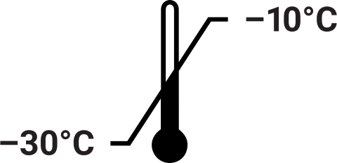
U.S. Pat. No. 8,809,529, European Pat. No. 2635582, Japanese Pat. No. 5889910 and other patents and patents pending.
U.S. Pat. Nos. 9,797,889, 9,797,890, 10,107,800 and 11,493,504; European Pat. No. 2970412; Japanese Pat. Nos. 7280842 and 7532562; and other patents and patents pending.
選択製品の構成品内容
| Item | Part # | Size |
|---|---|---|
Lumit® Detection Substrate B |
VB405A | 5 × 160μl |
Lumit® Detection Buffer B |
VB406A | 5 × 3.2ml |
Lumit® Antibody Dilution Buffer B |
W150A | 5 × 5.5ml |
Lumit® Anti-Insulin mAb-SmBiT, 200X |
W152A | 5 × 30μl |
Lumit® Anti-Insulin mAb-LgBiT, 200X |
W153A | 5 × 30μl |
U.S. Pat. No. 8,809,529, European Pat. No. 2635582, Japanese Pat. No. 5889910 and other patents and patents pending.
U.S. Pat. Nos. 9,797,889, 9,797,890, 10,107,800 and 11,493,504; European Pat. No. 2970412; Japanese Pat. Nos. 7280842 and 7532562; and other patents and patents pending.
Resources
Featured Citation
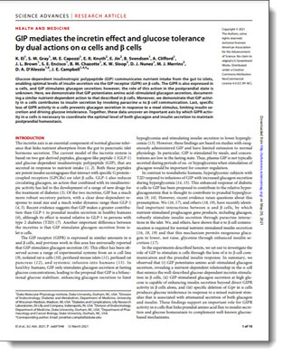
In this work, El and colleagues use the Lumit® Glucagon Immunoassay to characterize the complex regulatory mechanisms that control glucagon secretion from alpha cells.
El, K. et al. (2021) GIP mediates the incretin effect and glucose tolerance by dual actions on α cells and β cells. Sci. Adv. 7, abf1948.
技術記事
- Beyond Viability: Functional Profiling in Perifusion-Based Islet Studies
- Understanding the Complicated Relationship Between Insulin, Glucagon and GIP
- Blog Article: Jon Campbell Is Challenging Classic Models of Metabolic Disease
- Blog Article: Infographic: Assays for Measuring Insulin Activity
- White Paper: GLP-1 Signaling in Obesity Technologies for Monitoring GPCR Dynamics
- Application Note: Measuring Insulin Release Following GLP-1 Receptor Agonist Treatment using the Lumit® Insulin Immunoassay
技術ポスター
Related Products
類似製品
Lumit® Glucagon Immunoassay
迅速かつ簡単な洗浄不要プロトコルを用いて、細胞培養液や膵島分泌サンプル中のグルカゴンを定量的に測定
W8020, W8022
Lumit® IL-1β Human/Mouse Immunoassay
シンプルな無洗浄プロトコルを使用して、細胞培養サンプル中に放出されたIL-1βを定量的に測定。
W6010, W6011, W6012, W7010, W7011, W7012, W116A-C, W119A-C
Lumit® TNF-α (Human) Immunoassay
Quantitatively measures released TNF-α in cell culture samples using a simple, no-wash protocol.
W6050, W6052, W6051, W137A-C
Lumit® IL-6 (Human) Immunoassay
Quantitatively measures released IL-6 in cell culture samples using a simple, no-wash protocol.
W6030, W6032, W6031, W128A-C
関連製品
Glucose Uptake-Glo™ Assay
2DG6P の検出をベースにした哺乳動物細胞でのNon-RI グルコース取込み量を測定する発光アッセイ
J1341, J1342, J1343
Triglyceride-Glo™ Assay
リパーゼによる酵素反応で放出されるグリセロールの測定によりトリグリセリドを検出
J3160, J3161
Glycogen-Glo™ Assay
Fast, sensitive method for detecting glycogen in biological samples.
J5051, J5052
Incretin-Related Bioassays
Metabolic disease bioassays designed for potency testing and functional analysis.
CS3629B22, CS3629B24, CS3629B26, CS3629B15, CS3629B17, CS3629B19, CS3629B05, CS3629B07, CS3629B02, CS354805, CS354807, CS354802