Lumit® Cell Proliferation Assay (Human Ki-67)
Ki-67 検出のためのウォッシュ不要アッセイ
- 細胞増殖の代表的マーカーを用いたデータ信頼性の向上
- 洗浄不要・迅速なアッセイプロトコルによる前処理時間の短縮
- 増殖の早期指標に基づく迅速な意思決定

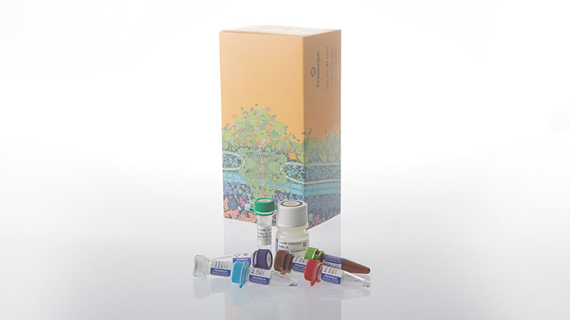
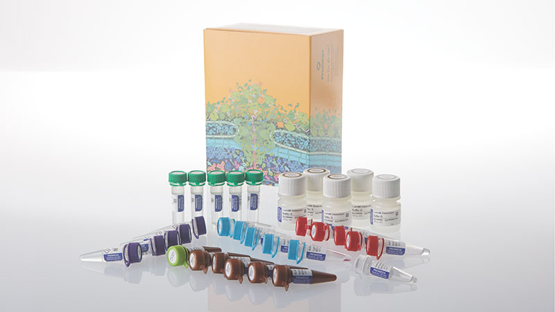
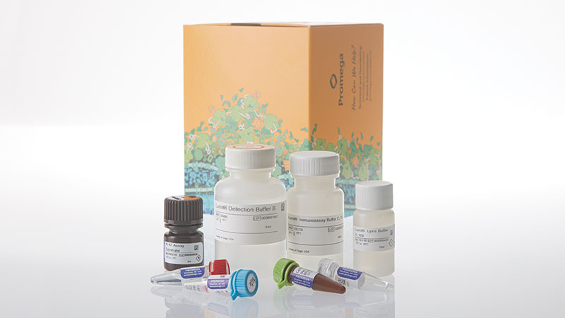
Catalog Number:
Size
Catalog Number: GC1000
Catalog Number: GC1002
Catalog Number: GC1001
「添加・混合・測定」アッセイにより、2時間以内に細胞増殖を評価
細胞増殖は、さまざまなアプリケーションワークフローで評価される、細胞ベース研究の基本的なパラメータです。現在、研究者は煩雑で信頼性に課題のある方法で細胞増殖をモニタリングしており、多くの場合、72~96時間の処理時間を必要とします。
Lumit® Cell Proliferation Assay(Human Ki-67)は、複雑な洗浄ステップを必要とせず、細胞増殖マーカーとして広く知られるhKi-67を追跡できる、シンプルな添加・測定型のプレートベースアッセイです。より早い時点で検出可能な堅牢なレスポンスにより、研究者はサンプル調製の手間と結果取得までの時間を削減しながら、より多くのデータを高い信頼性で取得できます。
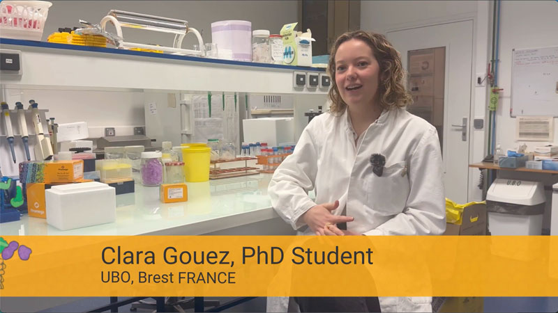
Clara Gouez氏が、Lumit® Cell Proliferation Assay(Human Ki-67)を胃がん研究の進展にどのように活用しているかを、動画でご覧ください。
Lumit® Cell Proliferation Assay の仕組み

シンプルなワンプレートプロトコル、洗浄ステップなし!

従来の固定処理を伴うKi-67フローサイトメトリーと比較して、このアッセイは大幅にシンプルかつ迅速で、解離や洗浄のステップを必要とせず、堅牢で再現性の高い結果が得られます。
Zeynep Kaya博士、博士研究員、Andrew Beggs教授グループ、バーミンガム大学
Lumit® hKi-67検出の広いダイナミックレンジと特異性


細胞増殖の減少を測定


Jurkat細胞(10,000 cells/well、384ウェルプレート、標準96ウェルの1/4容量)の、濃度を段階的に上げた化合物での処理 両化合物はいずれも、hKi-67発現量を濃度依存的に低下させたが、BAY-1895344では細胞毒性が認められた。Palbociclibは、わずか24時間の処理で細胞毒性を引き起こすことなくhKi-67量を大きく変化させ、増殖に対する化合物の影響をより早期に評価できることを示した。総ATP量に基づく評価では、Palbociclibの影響は小さいように見える。
細胞増殖の増加を測定


3D培養法に対応

3Dスフェロイドでは、hKi-67タンパク質量の低下により、生細胞数の代謝指標よりも広いレスポンスウィンドウで、抗増殖活性を明確かつ早期に確認できる HCT116細胞(1,000 cells/well)をSBio PrimeSurface 96Uプレート(ULA、丸底)に播種し、3日間培養して3Dスフェロイドを形成した。得られたHCT116スフェロイドを、濃度を段階的に上げたnutlin-3aで24時間処理した。その後、別々のプレートでLumit® Cell Proliferation Assay(Human Ki-67)および代謝活性アッセイを用いて増殖を評価した。 注:4日目の未処理HCT116スフェロイドの直径は約440µmであった。
プロトコル
製品仕様
Catalog Number:
選択製品の構成品内容
| Item | Part # | Size |
|---|---|---|
CellTox™ Green Dye, 1,000X |
G873A | 1 × 20μl |
Human Ki-67 Protein (Partial) Positive Control |
GC100A | 1 × 25μl |
Anti-hKi-67 mAb SmBiT, 400X |
GC101A | 1 × 60μl |
Anti-hKi-67 mAb LgBiT, 400X |
GC102A | 1 × 60μl |
Lumit® Immunoassay Buffer C, 10X |
VB115C | 1 × 1.8ml |
Lumit® Lysis Buffer II, 10X |
VB310C | 1 × 1.3ml |
Ki-67 Assay Substrate |
VB321A | 1 × 600μl |
Lumit® Detection Buffer B |
VB406D | 1 × 6ml |
U.S. Pat. No. 8,809,529, European Pat. No. 2635582, Japanese Pat. No. 5889910 and other patents and patents pending.
U.S. Pat. Nos. 9,797,889, 9,797,890, 10,107,800 and 11,493,504; European Pat. No. 2970412; Japanese Pat. Nos. 7280842 and 7532562; and other patents and patents pending.
選択製品の構成品内容
| Item | Part # | Size |
|---|---|---|
CellTox™ Green Dye, 1,000X |
G873B | 1 × 200μl |
Human Ki-67 Protein (Partial) Positive Control |
GC100A | 1 × 25μl |
Anti-hKi-67 mAb SmBiT, 400X |
GC101A | 5 × 60μl |
Anti-hKi-67 mAb LgBiT, 400X |
GC102A | 5 × 60μl |
Lumit® Immunoassay Buffer C, 10X |
VB115C | 5 × 1.8ml |
Lumit® Lysis Buffer II, 10X |
VB310C | 5 × 1.3ml |
Ki-67 Assay Substrate |
VB321A | 5 × 600μl |
Lumit® Detection Buffer B |
VB406D | 5 × 6ml |
U.S. Pat. No. 8,809,529, European Pat. No. 2635582, Japanese Pat. No. 5889910 and other patents and patents pending.
U.S. Pat. Nos. 9,797,889, 9,797,890, 10,107,800 and 11,493,504; European Pat. No. 2970412; Japanese Pat. Nos. 7280842 and 7532562; and other patents and patents pending.
選択製品の構成品内容
| Item | Part # | Size |
|---|---|---|
CellTox™ Green Dye, 1,000X |
G873B | 1 × 200μl |
Human Ki-67 Protein (Partial) Positive Control |
GC100A | 1 × 25μl |
Anti-hKi-67 mAb SmBiT, 400X |
GC101B | 1 × 600μl |
Anti-hKi-67 mAb LgBiT, 400X |
GC102B | 1 × 600μl |
Lumit® Immunoassay Buffer C, 10X |
VB115D | 1 × 18ml |
Lumit® Lysis Buffer II, 10X |
VB310D | 1 × 13ml |
Ki-67 Assay Substrate |
VB321B | 1 × 6ml |
Lumit® Detection Buffer B |
VB406E | 1 × 60ml |
U.S. Pat. No. 8,809,529, European Pat. No. 2635582, Japanese Pat. No. 5889910 and other patents and patents pending.
U.S. Pat. Nos. 9,797,889, 9,797,890, 10,107,800 and 11,493,504; European Pat. No. 2970412; Japanese Pat. Nos. 7280842 and 7532562; and other patents and patents pending.
技術資料
注目の資料
抗増殖作用と細胞死を区別する必要がありますか?
細胞増殖の主要マーカーであるhKi-67を検出するホモジニアスな細胞ベースアッセイ、Lumit® Cell Proliferation Assay(Human Ki-67)を用いることで、細胞増殖をすばやく簡単に評価できます。
技術記事
技術ポスター
- Poster: Homogeneous Bioluminescent Immunoassay for hKi-67: A Simple and Robust Screening Tool for Antiproliferative Agents
- Poster: Novel, Luminescent Ki-67 Immunoassay Assesses T Cell Responses
- Poster: Lumit® Immunoassays: Bioluminescent, Sensitive, and Homogeneous Analyte Detection Using Labeled Antibodies
製品比較
|
Cell Viability (ATP) |
Cell Viability (ATP) |
3D Cell Viability (ATP) |
Real-Time Viability |
Non-Lytic Viability |
Cell Proliferation |
|
|---|---|---|---|---|---|---|
| Best Use | Routine viability & HTS screening | Established protocols using original formulation | 3D spheroids, organoids, microtissues | Kinetic monitoring over time; downstream multiplexing | Multiplex first step; cells needed for follow-up assays | True proliferation readout independent of metabolism |
| Key Decision Points | ||||||
| Measures | Viability | Viability | Viability (3D) | Viability (kinetic) | Viability | Proliferation |
| Cells alive after assay? | ✗ Lytic | ✗ Lytic | ✗ Lytic | ✓ Non-lytic | ✓ Non-lytic | ✗ Lytic |
| Multiplexing compatible? | LimitedLytic—must be terminal step | LimitedLytic—must be terminal step | LimitedLytic—must be terminal step | ✓ ExcellentNon-lytic; pair with any downstream assay | ✓ ExcellentNon-lytic; pair with Caspase-Glo®, CTG, etc. | ModerateCan multiplex with CellTox™ Green or other fluorescent readouts |
| Real-time monitoring? | ✗ Endpoint | ✗ Endpoint | ✗ Endpoint | ✓ Up to 72hRead same wells repeatedly | ✗ Endpoint | ✗ Endpoint |
| 3D culture compatible? | PartialWorks for small spheroids; use 3D version for dense structures | PartialSame as 2.0 | ✓ OptimizedEnhanced lysis for dense 3D structures | PartialSubstrate must penetrate; best for small/loose 3D models | PartialSubstrate access may be limited in dense 3D | ✓ YesDetects Ki-67 in cell lysates from any culture format |
| Assay Attributes | ||||||
| Assay Principle | ATP quantitation (luciferase/luciferin) | ATP quantitation (luciferase/luciferin) | ATP quantitation (enhanced lysis for 3D) | Metabolic reduction of pro-substrate to luciferase substrate | Live-cell protease activity (GF-AFC cleavage) | Ki-67 immunodetection via NanoBiT® complementation |
| Detection Mode | Luminescence | Luminescence | Luminescence | Luminescence | Fluorescence400Ex / 505Em | Luminescence |
| Reagent Format | Ready-to-use liquid | Buffer + lyophilized substrateRequires reconstitution | Ready-to-use liquid | 2 components(enzyme + substrate) | Single reagent | Antibody mix + detection reagent |
| Time to Result | 10min | 10min | ~30min | ContinuousFirst read: 1–2h after addition | 30min | ~2h |
| Practical Considerations | ||||||
| Plate Formats | 96, 384, 1536 | 96, 384, 1536 | 96, 384 | 96, 384 | 96, 384 | 96, 384 |
| HTS Suitability | ✓ Excellent1536-well capable; fast protocol | ✓ Excellent1536-well capable | ✓ Good | ModerateRequires kinetic reader scheduling | ✓ Good | ✓ Good |
| Sensitivity (96-well) | ~15 cells/well | ~10 cells/well | Spheroid-dependent | <100 cells/well | ~40 cells/well | Cell line-dependent |
類似製品
CellTiter-Glo® Luminescent Cell Viability Assay
ATP定量に基づく培養細胞の生存数測定
G7570, G7571, G7572, G7573
CellTiter-Glo® 2.0 Cell Viability Assay
試薬の安定性が向上したCellTiter-Glo®、ATP検出に基づく細胞増殖の定量が可能
G9241, G9242, G9243
RealTime-Glo™ MT Cell Viability Assay
72 時間まで培養細胞の生存性をモニタリングできる発光アッセイ
G9711, G9712, G9713
CellTiter 96® AQueous One Solution Cell Proliferation Assay
細胞増殖、細胞毒性あるいは化学物質感受性試験などで生細胞数を測定するための発色アッセイ法
G3582, G3580, G3581